6VS8 | pdb_00006vs8

Mycobacterium tuberculosis dihydrofolate reductase in complex with ethyl 2-methyl thiazole-4-carboxylate(fragment 3)


Experimental Data Snapshot

  • Method: X-RAY DIFFRACTION
  • Resolution: 1.83 Å
  • R-Value Free: 
    0.221 (Depositor), 0.222 (DCC) 
  • R-Value Work: 
    0.170 (Depositor), 0.170 (DCC) 
  • R-Value Observed: 
    0.172 (Depositor) 

Starting Model: experimental
View more details

wwPDB Validation 3D Report Full Report

Validation slider image for 6VS8

Ligand Structure Quality Assessment 


This is version 1.2 of the entry. See complete history

Literature

Using a Fragment-Based Approach to Identify Alternative Chemical Scaffolds Targeting Dihydrofolate Reductase fromMycobacterium tuberculosis.

Ribeiro, J.A.Hammer, A.Libreros-Zuniga, G.A.Chavez-Pacheco, S.M.Tyrakis, P.de Oliveira, G.S.Kirkman, T.El Bakali, J.Rocco, S.A.Sforca, M.L.Parise-Filho, R.Coyne, A.G.Blundell, T.L.Abell, C.Dias, M.V.B.

(2020) ACS Infect Dis 6: 2192-2201

  • DOI: https://doi.org/10.1021/acsinfecdis.0c00263
  • Primary Citation Related Structures: 
    6VS5, 6VS6, 6VS8, 6VS9, 6VSD, 6VSE, 6VSF, 6VSG, 6VV6, 6VV7, 6VV8, 6VV9, 6VVB

  • PubMed Abstract: 

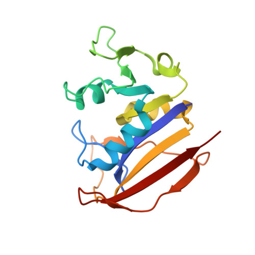
    Dihydrofolate reductase (DHFR), a key enzyme involved in folate metabolism, is a widely explored target in the treatment of cancer, immune diseases, bacteria, and protozoa infections. Although several antifolates have proved successful in the treatment of infectious diseases, they have been underexplored to combat tuberculosis, despite the essentiality of M. tuberculosis DHFR (MtDHFR). Herein, we describe an integrated fragment-based drug discovery approach to target MtDHFR that has identified hits with scaffolds not yet explored in any previous drug design campaign for this enzyme. The application of a SAR by catalog strategy of an in house library for one of the identified fragments has led to a series of molecules that bind to MtDHFR with low micromolar affinities. Crystal structures of MtDHFR in complex with compounds of this series demonstrated a novel binding mode that considerably differs from other DHFR antifolates, thus opening perspectives for the development of relevant MtDHFR inhibitors.


  • Organizational Affiliation
    • Department of Microbiology, Institute of Biomedical Science, University of São Paulo, Av. Prof. Lineu Prestes, 1474, São Paulo, SP 05508-000, Brazil.

Macromolecule Content 

  • Total Structure Weight: 38.4 kDa 
  • Atom Count: 2,982 
  • Modeled Residue Count: 318 
  • Deposited Residue Count: 322 
  • Unique protein chains: 1

Macromolecules

Find similar proteins by:|  3D Structure
Entity ID: 1
MoleculeChains  Sequence LengthOrganismDetailsImage
Dihydrofolate reductase
A, B
161Mycobacterium tuberculosisMutation(s): 0 
Gene Names: folAERS023446_01268ERS027651_02380SAMEA2682864_02011SAMEA2683035_01637
EC: 1.5.1.3
UniProt
Find proteins for P9WNX1 (Mycobacterium tuberculosis (strain ATCC 25618 / H37Rv))
Explore P9WNX1 
Go to UniProtKB:  P9WNX1
Entity Groups
Sequence Clusters30% Identity50% Identity70% Identity90% Identity95% Identity100% Identity
UniProt GroupP9WNX1
Sequence Annotations
Expand
Reference Sequence

Small Molecules

Ligands 5 Unique
IDChains Name / Formula / InChI Key2D Diagram3D Interactions
NAP

Query on NAP


Download:Ideal Coordinates CCD File
D [auth A],
I [auth B]
NADP NICOTINAMIDE-ADENINE-DINUCLEOTIDE PHOSPHATE
C21 H28 N7 O17 P3
XJLXINKUBYWONI-NNYOXOHSSA-N
RJY
(Subject of Investigation/LOI)

Query on RJY


Download:Ideal Coordinates CCD File
F [auth A],
L [auth B]
ethyl 2-methyl-1,3-thiazole-4-carboxylate
C7 H9 N O2 S
QWWPUBQHZFHZSF-UHFFFAOYSA-N
SO4

Query on SO4


Download:Ideal Coordinates CCD File
G [auth A],
M [auth B],
N [auth B],
O [auth B]
SULFATE ION
O4 S
QAOWNCQODCNURD-UHFFFAOYSA-L
PO4

Query on PO4


Download:Ideal Coordinates CCD File
E [auth A],
J [auth B],
K [auth B]
PHOSPHATE ION
O4 P
NBIIXXVUZAFLBC-UHFFFAOYSA-K
CO

Query on CO


Download:Ideal Coordinates CCD File
C [auth A],
H [auth B]
COBALT (II) ION
Co
XLJKHNWPARRRJB-UHFFFAOYSA-N

Experimental Data & Validation

Experimental Data

  • Method: X-RAY DIFFRACTION
  • Resolution: 1.83 Å
  • R-Value Free:  0.221 (Depositor), 0.222 (DCC) 
  • R-Value Work:  0.170 (Depositor), 0.170 (DCC) 
  • R-Value Observed: 0.172 (Depositor) 
Space Group: P 21 21 21
Unit Cell:
Length ( Å )Angle ( ˚ )
a = 60.278α = 90
b = 70.779β = 90
c = 72.108γ = 90
Software Package:
Software NamePurpose
Aimlessdata scaling
PHENIXrefinement
PDB_EXTRACTdata extraction
XDSdata reduction
PHASERphasing

Structure Validation

View Full Validation Report


Ligand Structure Quality Assessment 


Entry History 

& Funding Information

Deposition Data


Funding OrganizationLocationGrant Number
Sao Paulo Research Foundation (FAPESP)Brazil2018/00351-1

Revision History  (Full details and data files)

  • Version 1.0: 2020-07-15
    Type: Initial release
  • Version 1.1: 2020-08-26
    Changes: Database references, Derived calculations
  • Version 1.2: 2023-10-11
    Changes: Data collection, Database references, Refinement description