6O44 | pdb_00006o44

Insight into subtilisin E-S7 cleavage pattern based on crystal structure and hydrolysates peptide analysis

  • Classification: HYDROLASE
  • Organism(s): Bacillus subtilis
  • Expression System: Escherichia coli
  • Mutation(s): Yes 

  • Deposited: 2019-02-28 Released: 2019-04-10 
  • Deposition Author(s): Tang, H., Shi, K., Aihara, H.
  • Funding Organization(s): National Institutes of Health/National Institute of General Medical Sciences (NIH/NIGMS), National Natural Science Foundation of China (NSFC)

Experimental Data Snapshot

  • Method: X-RAY DIFFRACTION
  • Resolution: 1.83 Å
  • R-Value Free: 
    0.222 (Depositor), 0.222 (DCC) 
  • R-Value Work: 
    0.187 (Depositor), 0.191 (DCC) 
  • R-Value Observed: 
    0.189 (Depositor) 

Starting Model: experimental
View more details

wwPDB Validation 3D Report Full Report

Validation slider image for 6O44

This is version 1.3 of the entry. See complete history

Literature

Insight into subtilisin E-S7 cleavage pattern based on crystal structure and hydrolysates peptide analysis.

Tang, H.Zhang, J.Shi, K.Aihara, H.Du, G.

(2019) Biochem Biophys Res Commun 512: 623-628

  • DOI: https://doi.org/10.1016/j.bbrc.2019.03.064
  • Primary Citation Related Structures: 
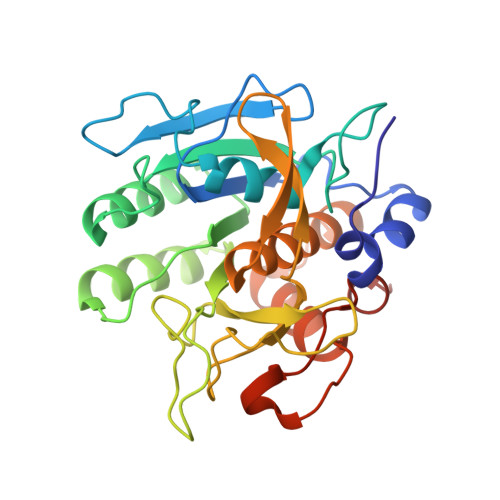
    6O44

  • PubMed Abstract: 

    The X-ray crystallographic structure of the mature form of subtilisin E-S7 (SES7) at 1.90 Å resolution is reported here. Structural comparisons between the previously reported propeptide-subtilisin E complex (1SCJ) and our mature form subtilisin E-S7 (6O44) provide insight into active site adjustments involved in catalysis and specificity. To further investigate the protease substrate selectivity mechanism, we used SES7 to hydrolyze skim milk and analyzed the hydrolysates by LC-MS for peptide identification. The cleavage pattern suggests a high preference for proline at substrate P2 position. The results based on the peptide analysis are consistent with our structural observations, which is instrumental in future protein engineering by rational design. Furthermore, the ACE-inhibitor and NLN-inhibitor activity of the hydrolysates were determined to assess the utility of SES7 for further industrial applications; IC 50 -ACE = 67 ± 0.92 μg/mL and IC 50 -NLN = 263 ± 13 μg/mL.


  • Organizational Affiliation
    • Key Laboratory of Industrial Biotechnology, Ministry of Education, School of Biotechnology, Jiangnan University, 1800 Lihu Road, Wuxi, 214122, Jiangsu, China; School of Biotechnology, Jiangnan University, 1800 Lihu Road, Wuxi, 214122, Jiangsu, China.

Macromolecule Content 

  • Total Structure Weight: 58.35 kDa 
  • Atom Count: 4,405 
  • Modeled Residue Count: 552 
  • Deposited Residue Count: 568 
  • Unique protein chains: 1

Macromolecules

Find similar proteins by:|  3D Structure
Entity ID: 1
MoleculeChains  Sequence LengthOrganismDetailsImage
Nattokinase
A, B
284Bacillus subtilisMutation(s): 1 
Gene Names: aprN
EC: 3.4.21.62
UniProt
Find proteins for P04189 (Bacillus subtilis (strain 168))
Explore P04189 
Go to UniProtKB:  P04189
Entity Groups
Sequence Clusters30% Identity50% Identity70% Identity90% Identity95% Identity100% Identity
UniProt GroupP04189
Sequence Annotations
Expand
Reference Sequence

Small Molecules

Ligands 3 Unique
IDChains Name / Formula / InChI Key2D Diagram3D Interactions
SO4

Query on SO4


Download:Ideal Coordinates CCD File
E [auth A]SULFATE ION
O4 S
QAOWNCQODCNURD-UHFFFAOYSA-L
EDO

Query on EDO


Download:Ideal Coordinates CCD File
F [auth A]
G [auth A]
H [auth A]
I [auth A]
L [auth B]
F [auth A],
G [auth A],
H [auth A],
I [auth A],
L [auth B],
M [auth B]
1,2-ETHANEDIOL
C2 H6 O2
LYCAIKOWRPUZTN-UHFFFAOYSA-N
CA

Query on CA


Download:Ideal Coordinates CCD File
C [auth A],
D [auth A],
J [auth B],
K [auth B]
CALCIUM ION
Ca
BHPQYMZQTOCNFJ-UHFFFAOYSA-N

Experimental Data & Validation

Experimental Data

  • Method: X-RAY DIFFRACTION
  • Resolution: 1.83 Å
  • R-Value Free:  0.222 (Depositor), 0.222 (DCC) 
  • R-Value Work:  0.187 (Depositor), 0.191 (DCC) 
  • R-Value Observed: 0.189 (Depositor) 
Space Group: P 21 21 21
Unit Cell:
Length ( Å )Angle ( ˚ )
a = 74.177α = 90
b = 80.123β = 90
c = 87.729γ = 90
Software Package:
Software NamePurpose
PHENIXrefinement
XDSdata reduction
Aimlessdata scaling
PHASERphasing

Structure Validation

View Full Validation Report


Entry History 

& Funding Information

Deposition Data


Funding OrganizationLocationGrant Number
National Institutes of Health/National Institute of General Medical Sciences (NIH/NIGMS)United StatesGM118047
National Natural Science Foundation of China (NSFC)China31470160

Revision History  (Full details and data files)

  • Version 1.0: 2019-04-10
    Type: Initial release
  • Version 1.1: 2019-05-01
    Changes: Data collection, Database references
  • Version 1.2: 2020-01-01
    Changes: Author supporting evidence
  • Version 1.3: 2023-10-11
    Changes: Data collection, Database references, Derived calculations, Refinement description