6HSW

A CE15 glucuronoyl esterase from Teredinibacter turnerae T7901


Experimental Data Snapshot

  • Method: X-RAY DIFFRACTION
  • Resolution: 2.15 Å
  • R-Value Free: 0.212 
  • R-Value Work: 0.164 
  • R-Value Observed: 0.164 

wwPDB Validation   3D Report Full Report


This is version 1.1 of the entry. See complete history


Literature

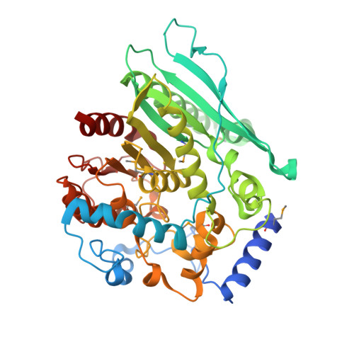
Structure-function analyses reveal that a glucuronoyl esterase fromTeredinibacter turneraeinteracts with carbohydrates and aromatic compounds.

Arnling Baath, J.Mazurkewich, S.Poulsen, J.N.Olsson, L.Lo Leggio, L.Larsbrink, J.

(2019) J Biol Chem 294: 6635-6644

  • DOI: https://doi.org/10.1074/jbc.RA119.007831
  • Primary Citation of Related Structures:  
    6HSW

  • PubMed Abstract: 

    Glucuronoyl esterases (GEs) catalyze the cleavage of ester linkages found between lignin and glucuronic acid moieties on glucuronoxylan in plant biomass. As such, GEs represent promising biochemical tools in industrial processing of these recalcitrant resources. However, details on how GEs interact with their natural substrates are sparse, calling for thorough structure-function studies. Presented here is the structure and biochemical characterization of a GE, Tt CE15A, from the bacterium Teredinibacter turnerae , a symbiont of wood-boring shipworms. To gain deeper insight into enzyme-substrate interactions, inhibition studies were performed with both the WT Tt CE15A and variants in which we, by using site-directed mutagenesis, substituted residues suggested to have key roles in binding to or interacting with the aromatic and carbohydrate structures of its uronic acid ester substrates. Our results support the hypothesis that two aromatic residues (Phe-174 and Trp-376), conserved in bacterial GEs, interact with aromatic and carbohydrate structures of these substrates in the enzyme active site, respectively. The solved crystal structure of Tt CE15A revealed features previously not observed in either fungal or bacterial GEs, with a large inserted N-terminal region neighboring the active site and a differently positioned residue of the catalytic triad. The findings highlight key interactions between GEs and complex lignin-carbohydrate ester substrates and advance our understanding of the substrate specificities of these enzymes in biomass conversion.


  • Organizational Affiliation

    From the Wallenberg Wood Science Center, Division of Industrial Biotechnology, Department of Biology and Biological Engineering, Chalmers University of Technology, SE-412 96 Gothenburg, Sweden and.


Macromolecules
Find similar proteins by:  (by identity cutoff)  |  3D Structure
Entity ID: 1
MoleculeChains Sequence LengthOrganismDetailsImage
Carbohydrate esterase family 15 domain protein
A, B, C
440Teredinibacter turnerae T7901Mutation(s): 0 
Gene Names: TERTU_0517
UniProt
Find proteins for C5BN23 (Teredinibacter turnerae (strain ATCC 39867 / T7901))
Explore C5BN23 
Go to UniProtKB:  C5BN23
Entity Groups  
Sequence Clusters30% Identity50% Identity70% Identity90% Identity95% Identity100% Identity
UniProt GroupC5BN23
Sequence Annotations
Expand
  • Reference Sequence
Small Molecules
Ligands 5 Unique
IDChains Name / Formula / InChI Key2D Diagram3D Interactions
1PE
Query on 1PE

Download Ideal Coordinates CCD File 
D [auth A],
P [auth B]
PENTAETHYLENE GLYCOL
C10 H22 O6
JLFNLZLINWHATN-UHFFFAOYSA-N
PEG
Query on PEG

Download Ideal Coordinates CCD File 
HA [auth C]DI(HYDROXYETHYL)ETHER
C4 H10 O3
MTHSVFCYNBDYFN-UHFFFAOYSA-N
GOL
Query on GOL

Download Ideal Coordinates CCD File 
AA [auth B]
BA [auth B]
CA [auth B]
DA [auth B]
EA [auth B]
AA [auth B],
BA [auth B],
CA [auth B],
DA [auth B],
EA [auth B],
FA [auth B],
L [auth A],
M [auth A],
N [auth A],
OA [auth C],
X [auth B],
Y [auth B],
Z [auth B]
GLYCEROL
C3 H8 O3
PEDCQBHIVMGVHV-UHFFFAOYSA-N
BR
Query on BR

Download Ideal Coordinates CCD File 
GA [auth C],
O [auth B]
BROMIDE ION
Br
CPELXLSAUQHCOX-UHFFFAOYSA-M
EDO
Query on EDO

Download Ideal Coordinates CCD File 
E [auth A]
F [auth A]
G [auth A]
H [auth A]
I [auth A]
E [auth A],
F [auth A],
G [auth A],
H [auth A],
I [auth A],
IA [auth C],
J [auth A],
JA [auth C],
K [auth A],
KA [auth C],
LA [auth C],
MA [auth C],
NA [auth C],
Q [auth B],
R [auth B],
S [auth B],
T [auth B],
U [auth B],
V [auth B],
W [auth B]
1,2-ETHANEDIOL
C2 H6 O2
LYCAIKOWRPUZTN-UHFFFAOYSA-N
Modified Residues  1 Unique
IDChains TypeFormula2D DiagramParent
MSE
Query on MSE
A, B, C
L-PEPTIDE LINKINGC5 H11 N O2 SeMET
Experimental Data & Validation

Experimental Data

  • Method: X-RAY DIFFRACTION
  • Resolution: 2.15 Å
  • R-Value Free: 0.212 
  • R-Value Work: 0.164 
  • R-Value Observed: 0.164 
  • Space Group: P 31 2 1
Unit Cell:
Length ( Å )Angle ( ˚ )
a = 121.173α = 90
b = 121.173β = 90
c = 198.203γ = 120
Software Package:
Software NamePurpose
XDSdata reduction
XSCALEdata scaling
AutoSolphasing
PHENIXrefinement

Structure Validation

View Full Validation Report


Entry History & Funding Information

Deposition Data


Funding OrganizationLocationGrant Number
Knut and Alice Wallenberg FoundationSwedenWood Science Center
Novo Nordisk FoundationDenmarkNNF17OC0027698
European UnionDenmarkInterreg-programmet for Oresund-Kattegat-Skagerrak

Revision History  (Full details and data files)

  • Version 1.0: 2019-03-20
    Type: Initial release
  • Version 1.1: 2019-05-29
    Changes: Data collection, Database references