5ZXL

Structure of GldA from E.coli


Experimental Data Snapshot

  • Method: X-RAY DIFFRACTION
  • Resolution: 2.79 Å
  • R-Value Free: 0.204 
  • R-Value Work: 0.173 
  • R-Value Observed: 0.174 

wwPDB Validation   3D Report Full Report


This is version 1.1 of the entry. See complete history


Literature

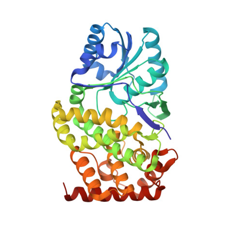
Structure of glycerol dehydrogenase (GldA) from Escherichia coli.

Zhang, J.Nanjaraj Urs, A.N.Lin, L.Zhou, Y.Hu, Y.Hua, G.Gao, Q.Yuchi, Z.Zhang, Y.

(2019) Acta Crystallogr F Struct Biol Commun 75: 176-183

  • DOI: https://doi.org/10.1107/S2053230X19000037
  • Primary Citation of Related Structures:  
    5ZXL

  • PubMed Abstract: 

    Escherichia coli (strain K-12, substrain MG1655) glycerol dehydrogenase (GldA) is required to catalyze the first step in fermentative glycerol metabolism. The protein was expressed and purified to homogeneity using a simple combination of heat-shock and chromatographic methods. The high yield of the protein (∼250 mg per litre of culture) allows large-scale production for potential industrial applications. Purified GldA exhibited a homogeneous tetrameric state (∼161 kDa) in solution and relatively high thermostability (T m = 65.6°C). Sitting-drop sparse-matrix screens were used for protein crystallization. An optimized condition with ammonium sulfate (2 M) provided crystals suitable for diffraction, and a binary structure containing glycerol in the active site was solved at 2.8 Å resolution. Each GldA monomer consists of nine β-strands, thirteen α-helices, two 3 10 -helices and several loops organized into two domains, the N- and C-terminal domains; the active site is located in a deep cleft between the two domains. The N-terminal domain contains a classic Rossmann fold for NAD + binding. The O 1 and O 2 atoms of glycerol serve as ligands for the tetrahedrally coordinated Zn 2+ ion. The orientation of the glycerol within the active site is mainly stabilized by van der Waals and electrostatic interactions with the benzyl ring of Phe245. Computer modeling suggests that the glycerol molecule is sandwiched by the Zn 2+ and NAD + ions. Based on this, the mechanism for the relaxed substrate specificity of this enzyme is also discussed.


  • Organizational Affiliation

    The Key Laboratory of Industrial Fermentation Microbiology, Ministry of Education, State Key Laboratory of Food Nutrition and Safety, Tianjin Engineering Research Center of Microbial Metabolism and Fermentation Process Control, College of Biotechnology, Tianjin University of Science and Technology, Tianjin 300457, People's Republic of China.


Macromolecules
Find similar proteins by:  (by identity cutoff)  |  3D Structure
Entity ID: 1
MoleculeChains Sequence LengthOrganismDetailsImage
Glycerol dehydrogenase
A, B, C, D
367Escherichia coli K-12Mutation(s): 0 
Gene Names: gldAb3945JW5556
EC: 1.1.1.6
UniProt
Find proteins for P0A9S5 (Escherichia coli (strain K12))
Explore P0A9S5 
Go to UniProtKB:  P0A9S5
Entity Groups  
Sequence Clusters30% Identity50% Identity70% Identity90% Identity95% Identity100% Identity
UniProt GroupP0A9S5
Sequence Annotations
Expand
  • Reference Sequence
Small Molecules
Ligands 4 Unique
IDChains Name / Formula / InChI Key2D Diagram3D Interactions
SO4
Query on SO4

Download Ideal Coordinates CCD File 
AA [auth D],
L [auth B],
R [auth C],
S [auth C],
Z [auth D]
SULFATE ION
O4 S
QAOWNCQODCNURD-UHFFFAOYSA-L
GOL
Query on GOL

Download Ideal Coordinates CCD File 
F [auth A]
G [auth A]
I [auth B]
J [auth B]
K [auth B]
F [auth A],
G [auth A],
I [auth B],
J [auth B],
K [auth B],
N [auth C],
O [auth C],
P [auth C],
Q [auth C],
V [auth D],
W [auth D],
X [auth D]
GLYCEROL
C3 H8 O3
PEDCQBHIVMGVHV-UHFFFAOYSA-N
ZN
Query on ZN

Download Ideal Coordinates CCD File 
E [auth A],
H [auth B],
M [auth C],
T [auth D],
U [auth D]
ZINC ION
Zn
PTFCDOFLOPIGGS-UHFFFAOYSA-N
CL
Query on CL

Download Ideal Coordinates CCD File 
Y [auth D]CHLORIDE ION
Cl
VEXZGXHMUGYJMC-UHFFFAOYSA-M
Experimental Data & Validation

Experimental Data

  • Method: X-RAY DIFFRACTION
  • Resolution: 2.79 Å
  • R-Value Free: 0.204 
  • R-Value Work: 0.173 
  • R-Value Observed: 0.174 
  • Space Group: P 43 21 2
Unit Cell:
Length ( Å )Angle ( ˚ )
a = 162.446α = 90
b = 162.446β = 90
c = 293.127γ = 90
Software Package:
Software NamePurpose
HKL-2000data scaling
PHENIXrefinement
PDB_EXTRACTdata extraction
HKL-3000data reduction
PHENIXphasing

Structure Validation

View Full Validation Report


Entry History 

Deposition Data

Revision History  (Full details and data files)

  • Version 1.0: 2019-03-20
    Type: Initial release
  • Version 1.1: 2023-11-22
    Changes: Data collection, Database references, Derived calculations, Refinement description