5ZVJ | pdb_00005zvj

Crystal structure of HtrA1 from Mycobacterium tuberculosis


Experimental Data Snapshot

  • Method: X-RAY DIFFRACTION
  • Resolution: 2.70 Å
  • R-Value Free: 
    0.250 (Depositor), 0.250 (DCC) 
  • R-Value Work: 
    0.195 (Depositor), 0.195 (DCC) 
  • R-Value Observed: 
    0.198 (Depositor) 

Starting Model: experimental
View more details

wwPDB Validation 3D Report Full Report

Validation slider image for 5ZVJ

This is version 1.4 of the entry. See complete history

Literature

The crystal structure of an essential high-temperature requirement protein HtrA1 (Rv1223) from Mycobacterium tuberculosis reveals its unique features.

Singh, K.H.Yadav, S.Kumar, D.Biswal, B.K.

(2018) Acta Crystallogr D Struct Biol 74: 906-921

  • DOI: https://doi.org/10.1107/S205979831800952X
  • Primary Citation Related Structures: 
    5ZVJ

  • PubMed Abstract: 

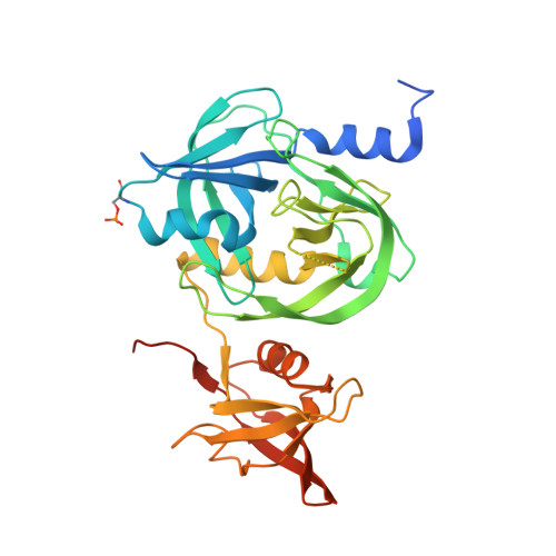
    High-temperature requirement A (HtrA) proteins, which are members of the heat-shock-induced serine protease family, are involved in extracytoplasmic protein quality control and bacterial survival strategies under stress conditions, and are associated with the virulence of several pathogens; they are therefore major drug targets. Mycobacterium tuberculosis possesses three putative HtrAs: HtrA1 (Rv1223), HtrA2 (Rv0983) and HtrA3 (Rv0125). Each has a cytoplasmic region, a transmembrane helix and a periplasmic region. Here, the crystal structure of the periplasmic region consisting of a protease domain (PD) and a PDZ domain from an M. tuberculosis HtrA1 mutant (mHtrA1 S387A ) is reported at 2.7 Å resolution. Although the mHtrA1 S387A PD shows structural features similar to those of other HtrAs, its loops, particularly L3 and LA, display different conformations. Loop L3 communicates between the PDs of the trimer and the PDZ domains and undergoes a transition from an active to an inactive conformation, as reported for an equivalent HtrA (DegS). Loop LA, which is responsible for higher oligomer formation owing to its length (50 amino acids) in DegP, is very short in mHtrA1 S387A (five amino acids), as in mHtrA2 (also five amino acids), and therefore lacks essential interactions for the formation of higher oligomers. Notably, a well ordered loop known as the insertion clamp in the PDZ domain interacts with the protease domain of the adjacent molecule, which possibly aids in the stabilization of a trimeric functional unit of this enzyme. The three-dimensional structure of mHtrA1 S387A presented here will be useful in the design of enzyme-specific antituberculosis inhibitors.


  • Organizational Affiliation
    • Structural and Functional Biology Laboratory, National Institute of Immunology, Aruna Asaf Ali Marg, New Delhi 110 067, India.

Macromolecule Content 

  • Total Structure Weight: 34.99 kDa 
  • Atom Count: 2,229 
  • Modeled Residue Count: 299 
  • Deposited Residue Count: 337 
  • Unique protein chains: 1

Macromolecules

Find similar proteins by:|  3D Structure
Entity ID: 1
MoleculeChains  Sequence LengthOrganismDetailsImage
Serine protease337Mycobacterium tuberculosisMutation(s): 1 
Gene Names: htrASAMEA2682835_04373
EC: 3.4.21 (PDB Primary Data), 3.4.21.107 (PDB Primary Data)
UniProt
Find proteins for O06291 (Mycobacterium tuberculosis (strain ATCC 25618 / H37Rv))
Explore O06291 
Go to UniProtKB:  O06291
Entity Groups
Sequence Clusters30% Identity50% Identity70% Identity90% Identity95% Identity100% Identity
UniProt GroupO06291
Sequence Annotations
Expand
Reference Sequence

Small Molecules

Ligands 1 Unique
IDChains Name / Formula / InChI Key2D Diagram3D Interactions
TRS

Query on TRS


Download:Ideal Coordinates CCD File
B [auth A]2-AMINO-2-HYDROXYMETHYL-PROPANE-1,3-DIOL
C4 H12 N O3
LENZDBCJOHFCAS-UHFFFAOYSA-O
Modified Residues  1 Unique
IDChains TypeFormula2D DiagramParent
SEP
Query on SEP
A
L-PEPTIDE LINKINGC3 H8 N O6 PSER

Experimental Data & Validation

Experimental Data

  • Method: X-RAY DIFFRACTION
  • Resolution: 2.70 Å
  • R-Value Free:  0.250 (Depositor), 0.250 (DCC) 
  • R-Value Work:  0.195 (Depositor), 0.195 (DCC) 
  • R-Value Observed: 0.198 (Depositor) 
Space Group: P 63
Unit Cell:
Length ( Å )Angle ( ˚ )
a = 135.391α = 90
b = 135.391β = 90
c = 45.852γ = 120
Software Package:
Software NamePurpose
REFMACrefinement
XDSdata reduction
XDSdata scaling
PHASERphasing

Structure Validation

View Full Validation Report


Entry History 

Deposition Data

Revision History  (Full details and data files)

  • Version 1.0: 2018-09-05
    Type: Initial release
  • Version 1.1: 2018-09-19
    Changes: Data collection, Database references
  • Version 1.2: 2019-12-25
    Changes: Database references
  • Version 1.3: 2023-11-22
    Changes: Data collection, Database references, Refinement description
  • Version 1.4: 2024-11-13
    Changes: Structure summary