5ZJ5 | pdb_00005zj5

Guanine-specific ADP-ribosyltransferase with NADH and GDP


Experimental Data Snapshot

  • Method: X-RAY DIFFRACTION
  • Resolution: 1.57 Å
  • R-Value Free: 
    0.208 (Depositor), 0.210 (DCC) 
  • R-Value Work: 
    0.174 (Depositor), 0.170 (DCC) 
  • R-Value Observed: 
    0.175 (Depositor) 

Starting Model: experimental
View more details

wwPDB Validation   3D Report Full Report


Ligand Structure Quality Assessment 


This is version 1.4 of the entry. See complete history


Literature

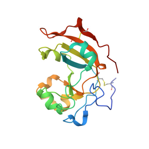
Substrate N2atom recognition mechanism in pierisin family DNA-targeting, guanine-specific ADP-ribosyltransferase ScARP.

Yoshida, T.Tsuge, H.

(2018) J Biological Chem 293: 13768-13774

  • DOI: https://doi.org/10.1074/jbc.AC118.004412
  • Primary Citation of Related Structures:  
    5ZJ4, 5ZJ5

  • PubMed Abstract: 

    ScARP from the bacterium Streptomyces coelicolor belongs to the pierisin family of DNA-targeting ADP-ribosyltransferases (ARTs). These enzymes ADP-ribosylate the N 2 amino groups of guanine residues in DNA to yield N 2 -(ADP-ribos-1-yl)-2'-deoxyguanosine. Although the structures of pierisin-1 and Scabin were revealed recently, the substrate recognition mechanisms remain poorly understood because of the lack of a substrate-binding structure. Here, we report the apo structure of ScARP and of ScARP bound to NADH and its GDP substrate at 1.50 and 1.57 Å resolutions, respectively. The bound structure revealed that the guanine of GDP is trapped between N -ribose of NADH and Trp-159. Interestingly, N 2 and N 3 of guanine formed hydrogen bonds with the OE1 and NE2 atoms of Gln-162, respectively. We directly observed that the ADP-ribosylating toxin turn-turn (ARTT)-loop, including Trp-159 and Gln-162, plays a key role in the specificity of DNA-targeting, guanine-specific ARTs as well as protein-targeting ARTs such as the C3 exoenzyme. We propose that the ARTT-loop recognition is a common substrate-recognition mechanism in the pierisin family. Furthermore, this complex structure sheds light on similarities and differences among two subclasses that are distinguished by conserved structural motifs: H-Y-E in the ARTD subfamily and R-S-E in the ARTC subfamily. The spatial arrangements of the electrophile and nucleophile were the same, providing the first evidence for a common reaction mechanism in these ARTs. ARTC (including ScARP) uses the ARTT-loop for substrate recognition, whereas ARTD (represented by Arr) uses the C-terminal helix instead of the ARTT-loop. These observations could help inform efforts to improve ART inhibitors.


  • Organizational Affiliation
    • From the Department of Bioresource and Environmental Sciences, Faculty of Life Sciences.

Macromolecules
Find similar proteins by:  (by identity cutoff)  |  3D Structure
Entity ID: 1
MoleculeChains Sequence LengthOrganismDetailsImage
ADP-ribosyltransferase
A, B
162Streptomyces coelicolor A3(2)Mutation(s): 0 
Gene Names: SCO5461
EC: 2.4.2
UniProt
Find proteins for Q9L1E4 (Streptomyces coelicolor (strain ATCC BAA-471 / A3(2) / M145))
Explore Q9L1E4 
Go to UniProtKB:  Q9L1E4
Entity Groups  
Sequence Clusters30% Identity50% Identity70% Identity90% Identity95% Identity100% Identity
UniProt GroupQ9L1E4
Sequence Annotations
Expand
  • Reference Sequence
Experimental Data & Validation

Experimental Data

  • Method: X-RAY DIFFRACTION
  • Resolution: 1.57 Å
  • R-Value Free:  0.208 (Depositor), 0.210 (DCC) 
  • R-Value Work:  0.174 (Depositor), 0.170 (DCC) 
  • R-Value Observed: 0.175 (Depositor) 
Space Group: C 1 2 1
Unit Cell:
Length ( Å )Angle ( ˚ )
a = 116.197α = 90
b = 40.049β = 113.967
c = 77.054γ = 90
Software Package:
Software NamePurpose
PHENIXrefinement
REFMACrefinement
HKL-2000data collection
HKL-2000data reduction
HKL-2000data scaling
PHASERphasing
Cootmodel building

Structure Validation

View Full Validation Report


Ligand Structure Quality Assessment 


Entry History & Funding Information

Deposition Data


Funding OrganizationLocationGrant Number
Japan Society for the Promotion of ScienceJapan17K15095
Japan Society for the Promotion of ScienceJapan15K08289
--

Revision History  (Full details and data files)

  • Version 1.0: 2018-08-08
    Type: Initial release
  • Version 1.1: 2018-09-05
    Changes: Data collection, Database references
  • Version 1.2: 2018-09-19
    Changes: Data collection, Database references
  • Version 1.3: 2023-11-22
    Changes: Data collection, Database references, Refinement description
  • Version 1.4: 2024-10-16
    Changes: Structure summary