5ZG6

Crystal structure of beta-lactamase PenP mutant-E166Y in complex with cephaloridine as "post-acylation" intermediate


Experimental Data Snapshot

  • Method: X-RAY DIFFRACTION
  • Resolution: 2.00 Å
  • R-Value Free: 0.258 
  • R-Value Work: 0.208 
  • R-Value Observed: 0.211 

wwPDB Validation   3D Report Full Report


Ligand Structure Quality Assessment 


This is version 1.2 of the entry. See complete history


Literature

The hydrolytic water molecule of Class A beta-lactamase relies on the acyl-enzyme intermediate ES* for proper coordination and catalysis.

He, Y.Lei, J.Pan, X.Huang, X.Zhao, Y.

(2020) Sci Rep 10: 10205-10205

  • DOI: https://doi.org/10.1038/s41598-020-66431-w
  • Primary Citation of Related Structures:  
    5ZFL, 5ZFT, 5ZG6

  • PubMed Abstract: 

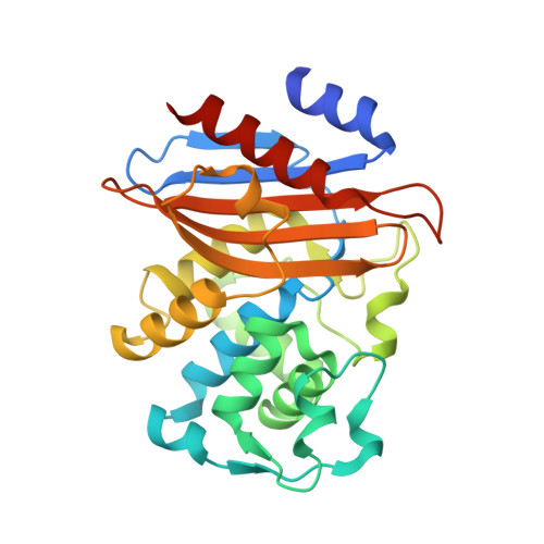
    Serine-based β-lactamases of Class A, C and D all rely on a key water molecule to hydrolyze and inactivate β-lactam antibiotics. This process involves two conserved catalytic steps. In the first acylation step, the β-lactam antibiotic forms an acyl-enzyme intermediate (ES*) with the catalytic serine residue. In the second deacylation step, an activated water molecule serves as nucleophile (WAT_Nu) to attack ES* and release the inactivated β-lactam. The coordination and activation of WAT_Nu is not fully understood. Using time-resolved x-ray crystallography and QM/MM simulations, we analyzed three intermediate structures of Class A β-lactamase PenP as it slowly hydrolyzed cephaloridine. WAT_Nu is centrally located in the apo structure but becomes slightly displaced away by ES* in the post-acylation structure. In the deacylation structure, WAT_Nu moves back and is positioned along the Bürgi-Dunitz trajectory with favorable energetic profile to attack ES*. Unexpectedly, WAT_Nu is also found to adopt a catalytically incompetent conformation in the deacylation structure forming a hydrogen bond with ES*. Our results reveal that ES* plays a significant role in coordinating and activating WAT_Nu through subtle yet distinct interactions at different stages of the catalytic process. These interactions may serve as potential targets to circumvent β-lactamase-mediated antibiotic resistance.


  • Organizational Affiliation

    The Hong Kong Polytechnic University Shenzhen Research Institute, Shenzhen, P. R. China.


Macromolecules
Find similar proteins by:  (by identity cutoff)  |  3D Structure
Entity ID: 1
MoleculeChains Sequence LengthOrganismDetailsImage
Beta-lactamaseA [auth B],
B [auth A]
268Bacillus licheniformisMutation(s): 1 
Gene Names: penPblaP
EC: 3.5.2.6
UniProt
Find proteins for P00808 (Bacillus licheniformis)
Explore P00808 
Go to UniProtKB:  P00808
Entity Groups  
Sequence Clusters30% Identity50% Identity70% Identity90% Identity95% Identity100% Identity
UniProt GroupP00808
Sequence Annotations
Expand
  • Reference Sequence
Small Molecules
Ligands 1 Unique
IDChains Name / Formula / InChI Key2D Diagram3D Interactions
CED
Query on CED

Download Ideal Coordinates CCD File 
C [auth B],
D [auth A]
5-METHYL-2-[2-OXO-1-(2-THIOPHEN-2-YL-ACETYLAMINO)-ETHYL]-3,6-DIHYDRO-2H-[1,3]THIAZINE-4-CARBOXYLIC ACID
C14 H16 N2 O4 S2
SFVACKBZMIZHCK-ZWNOBZJWSA-N
Experimental Data & Validation

Experimental Data

  • Method: X-RAY DIFFRACTION
  • Resolution: 2.00 Å
  • R-Value Free: 0.258 
  • R-Value Work: 0.208 
  • R-Value Observed: 0.211 
  • Space Group: P 1
Unit Cell:
Length ( Å )Angle ( ˚ )
a = 43.292α = 77.43
b = 46.459β = 75.53
c = 65.981γ = 69.34
Software Package:
Software NamePurpose
REFMACrefinement
SCALEPACKdata scaling
PDB_EXTRACTdata extraction
HKL-3000data reduction
PHASERphasing

Structure Validation

View Full Validation Report


Ligand Structure Quality Assessment 


Entry History 

Deposition Data

Revision History  (Full details and data files)

  • Version 1.0: 2019-03-20
    Type: Initial release
  • Version 1.1: 2020-07-08
    Changes: Database references
  • Version 1.2: 2023-11-22
    Changes: Data collection, Database references, Refinement description