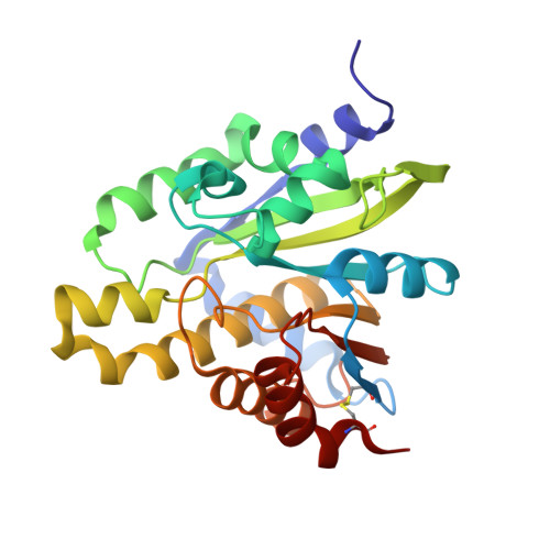
5ZDB

Crystal structure of poly(ADP-ribose) glycohydrolase (PARG) from Deinococcus radiodurans in complex with ADP-ribose (P21)


Experimental Data Snapshot

  • Method: X-RAY DIFFRACTION
  • Resolution: 1.97 Å
  • R-Value Free: 0.197 
  • R-Value Work: 0.155 
  • R-Value Observed: 0.158 

Starting Model: experimental
View more details

wwPDB Validation   3D Report Full Report


Ligand Structure Quality Assessment 


This is version 1.3 of the entry. See complete history


Literature

Structural and biochemical evidence supporting poly ADP-ribosylation in the bacterium Deinococcus radiodurans.

Cho, C.C.Chien, C.Y.Chiu, Y.C.Lin, M.H.Hsu, C.H.

(2019) Nat Commun 10: 1491-1491

  • DOI: https://doi.org/10.1038/s41467-019-09153-6
  • Primary Citation of Related Structures:  
    5ZDA, 5ZDB, 5ZDC, 5ZDD, 5ZDE, 5ZDF, 5ZDG

  • PubMed Abstract: 

    Poly-ADP-ribosylation, a post-translational modification involved in various cellular processes, is well characterized in eukaryotes but thought to be devoid in bacteria. Here, we solve crystal structures of ADP-ribose-bound poly(ADP-ribose)glycohydrolase from the radioresistant bacterium Deinococcus radiodurans (DrPARG), revealing a solvent-accessible 2'-hydroxy group of ADP-ribose, which suggests that DrPARG may possess endo-glycohydrolase activity toward poly-ADP-ribose (PAR). We confirm the existence of PAR in D. radiodurans and show that disruption of DrPARG expression causes accumulation of endogenous PAR and compromises recovery from UV radiation damage. Moreover, endogenous PAR levels in D. radiodurans are elevated after UV irradiation, indicating that PARylation may be involved in resistance to genotoxic stresses. These findings provide structural insights into a bacterial-type PARG and suggest the existence of a prokaryotic PARylation machinery that may be involved in stress responses.


  • Organizational Affiliation

    Genome and Systems Biology Degree Program, National Taiwan University and Academia Sinica, Taipei, 10617, Taiwan.


Macromolecules
Find similar proteins by:  (by identity cutoff)  |  3D Structure
Entity ID: 1
MoleculeChains Sequence LengthOrganismDetailsImage
Poly ADP-ribose glycohydrolase
A, B
253Deinococcus radiodurans R1 = ATCC 13939 = DSM 20539Mutation(s): 0 
Gene Names: DR_B0099
UniProt
Find proteins for Q9RZM4 (Deinococcus radiodurans (strain ATCC 13939 / DSM 20539 / JCM 16871 / CCUG 27074 / LMG 4051 / NBRC 15346 / NCIMB 9279 / VKM B-1422 / R1))
Explore Q9RZM4 
Go to UniProtKB:  Q9RZM4
Entity Groups  
Sequence Clusters30% Identity50% Identity70% Identity90% Identity95% Identity100% Identity
UniProt GroupQ9RZM4
Sequence Annotations
Expand
  • Reference Sequence
Small Molecules
Ligands 1 Unique
IDChains Name / Formula / InChI Key2D Diagram3D Interactions
AR6
Query on AR6

Download Ideal Coordinates CCD File 
C [auth A],
D [auth B]
[(2R,3S,4R,5R)-5-(6-AMINOPURIN-9-YL)-3,4-DIHYDROXY-OXOLAN-2-YL]METHYL [HYDROXY-[[(2R,3S,4R,5S)-3,4,5-TRIHYDROXYOXOLAN-2-YL]METHOXY]PHOSPHORYL] HYDROGEN PHOSPHATE
C15 H23 N5 O14 P2
SRNWOUGRCWSEMX-ZQSHOCFMSA-N
Experimental Data & Validation

Experimental Data

  • Method: X-RAY DIFFRACTION
  • Resolution: 1.97 Å
  • R-Value Free: 0.197 
  • R-Value Work: 0.155 
  • R-Value Observed: 0.158 
  • Space Group: P 1 21 1
Unit Cell:
Length ( Å )Angle ( ˚ )
a = 43.29α = 90
b = 97.427β = 93.65
c = 59.369γ = 90
Software Package:
Software NamePurpose
PHENIXrefinement
HKL-2000data reduction
HKL-2000data scaling
PHENIXphasing

Structure Validation

View Full Validation Report


Ligand Structure Quality Assessment 


Entry History & Funding Information

Deposition Data


Funding OrganizationLocationGrant Number
National Science Council (Taiwan)Taiwan105-2113-M-002-009

Revision History  (Full details and data files)

  • Version 1.0: 2019-02-27
    Type: Initial release
  • Version 1.1: 2020-03-11
    Changes: Database references, Structure summary
  • Version 1.2: 2023-11-22
    Changes: Data collection, Database references, Refinement description
  • Version 1.3: 2024-10-30
    Changes: Structure summary