Novel Mechanism for Cyclic Dinucleotide Degradation Revealed by Structural Studies of Vibrio Phosphodiesterase V-cGAP3.
Deng, M.J., Tao, J., E, C., Ye, Z.Y., Jiang, Z., Yu, J., Su, X.D.(2018) J Mol Biol 430: 5080-5093
- PubMed: 30365951 Search on PubMed
- DOI: https://doi.org/10.1016/j.jmb.2018.10.010
- Primary Citation Related Structures:
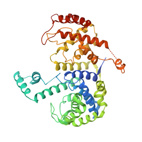
5Z7C - PubMed Abstract:
3'3'-cyclic GMP-AMP (3'3'-cGAMP) belongs to a family of the bacterial secondary messenger cyclic dinucleotides. It was first discovered in the Vibrio cholerae seventh pandemic strains and is involved in efficient intestinal colonization and chemotaxis regulation. Phosphodiesterases (PDEs) that degrade 3'3'-cGAMP play important regulatory roles in the relevant signaling pathways, and a previous study has identified three PDEs in V. cholerae, namely, V-cGAP1, V-cGAP2, and V-cGAP3, functioning in 3'3'-cGAMP degradation. We report the crystal structure, biochemical, and structural analyses of V-cGAP3, providing a foundation for understanding the mechanism of 3'3'-cGAMP degradation and regulation in general. Our crystal and molecular dynamic (MD)-simulated structures revealed that V-cGAP3 contains tandem HD-GYP domains within its N- and C-terminal domains, with similar three-dimensional topologies despite their low-sequence identity. Biochemical and structural analyses showed that the N-terminal domain plays a mechanism of positive regulation for the catalytic C-terminal domain. We also demonstrated that the other homologous Vibrio PDEs, V-cGAP1/2, likely function via a similar mechanism.
- State Key Laboratory of Protein and Plant Gene Research, and Biomedical Pioneering Innovation Center (BIOPIC), School of Life Sciences, Peking University, Beijing 100871, China.
Organizational Affiliation: