


Crystal structures of pyrrolidone carboxylate peptidase I from Deionococcus radiodurans reveal the mechanism of L-pyroglutamate recognition
Agrawal, R., Ghosh, B., Kumar, A., Makde, R.D.To be published.
Experimental Data Snapshot
Starting Model: experimental
View more details
wwPDB Validation 3D Report Full Report

Entity ID: 1 | |||||
|---|---|---|---|---|---|
| Molecule | Chains | Sequence Length | Organism | Details | Image |
| Pyrrolidone-carboxylate peptidase | 218 | Deinococcus radiodurans R1 = ATCC 13939 = DSM 20539 | Mutation(s): 0 Gene Names: pcp, DR_0490 EC: 3.4.19.3 | 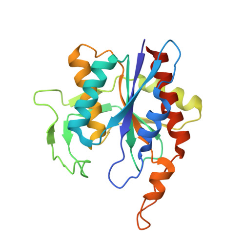 | |
UniProt | |||||
Entity Groups | |||||
| Sequence Clusters | 30% Identity50% Identity70% Identity90% Identity95% Identity100% Identity | ||||
| UniProt Group | Q9RX25 | ||||
Sequence AnnotationsExpand | |||||
Reference Sequence | |||||
| Length ( Å ) | Angle ( ˚ ) |
|---|---|
| a = 88.895 | α = 90 |
| b = 46.87 | β = 105.61 |
| c = 105.293 | γ = 90 |
| Software Name | Purpose |
|---|---|
| PHENIX | refinement |
| Coot | model building |
| PHENIX | model building |
| PHASER | phasing |
| Aimless | data scaling |
| XDS | data reduction |
| MAR345dtb | data collection |


