5YZ8 | pdb_00005yz8

Crystal Structure of N-terminal C1 domain of KaiC


Experimental Data Snapshot

  • Method: X-RAY DIFFRACTION
  • Resolution: 2.81 Å
  • R-Value Free: 
    0.300 (Depositor), 0.292 (DCC) 
  • R-Value Work: 
    0.241 (Depositor), 0.242 (DCC) 
  • R-Value Observed: 
    0.244 (Depositor) 

Starting Model: experimental
View more details

wwPDB Validation 3D Report Full Report

Validation slider image for 5YZ8

Ligand Structure Quality Assessment 


This is version 1.2 of the entry. See complete history

Literature

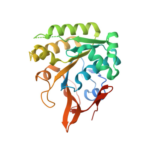
Conformational rearrangements of the C1 ring in KaiC measure the timing of assembly with KaiB.

Mukaiyama, A.Furuike, Y.Abe, J.Yamashita, E.Kondo, T.Akiyama, S.

(2018) Sci Rep 8: 8803-8803

  • DOI: https://doi.org/10.1038/s41598-018-27131-8
  • Primary Citation Related Structures: 
    5YZ8

  • PubMed Abstract: 

    KaiC, the core oscillator of the cyanobacterial circadian clock, is composed of an N-terminal C1 domain and a C-terminal C2 domain, and assembles into a double-ring hexamer upon ATP binding. Cyclic phosphorylation and dephosphorylation at Ser431 and Thr432 in the C2 domain proceed with a period of approximately 24 h in the presence of other clock proteins, KaiA and KaiB, but recent studies have revealed a crucial role for the C1 ring in determining the cycle period. In this study, we mapped dynamic structural changes of the C1 ring in solution using a combination of site-directed tryptophan mutagenesis and fluorescence spectroscopy. We found that the C1 ring undergoes a structural transition, coupled with ATPase activity and the phosphorylation state, while maintaining its hexameric ring structure. This transition triggered by ATP hydrolysis in the C1 ring in specific phosphorylation states is a necessary event for recruitment of KaiB, limiting the overall rate of slow complex formation. Our results provide structural and kinetic insights into the C1-ring rearrangements governing the slow dynamics of the cyanobacterial circadian clock.


  • Organizational Affiliation
    • Research Center of Integrative Molecular Systems (CIMoS), Institute for Molecular Science, National Institute for Natural Sciences, 38 Nishigo-Naka, Myodaiji, Okazaki, 444-8585, Japan. amukai@ims.ac.jp.

Macromolecule Content 

  • Total Structure Weight: 174.94 kDa 
  • Atom Count: 9,974 
  • Modeled Residue Count: 1,324 
  • Deposited Residue Count: 1,524 
  • Unique protein chains: 1

Macromolecules

Find similar proteins by:|  3D Structure
Entity ID: 1
MoleculeChains  Sequence LengthOrganismDetailsImage
Circadian Clock Protein Kinase KaiC
A, B, C, D, E
A, B, C, D, E, F
254Synechococcus elongatus PCC 7942 = FACHB-805Mutation(s): 1 
Gene Names: KAICSYNPCC7942_1216SEE0011
EC: 2.7.11.1 (PDB Primary Data), 3.6.4 (UniProt)
UniProt
Find proteins for Q79PF4 (Synechococcus elongatus (strain ATCC 33912 / PCC 7942 / FACHB-805))
Explore Q79PF4 
Go to UniProtKB:  Q79PF4
Entity Groups
Sequence Clusters30% Identity50% Identity70% Identity90% Identity95% Identity100% Identity
UniProt GroupQ79PF4
Sequence Annotations
Expand
Reference Sequence

Small Molecules

Ligands 3 Unique
IDChains Name / Formula / InChI Key2D Diagram3D Interactions
ANP

Query on ANP


Download:Ideal Coordinates CCD File
G [auth A]
K [auth B]
N [auth C]
Q [auth D]
T [auth E]
G [auth A],
K [auth B],
N [auth C],
Q [auth D],
T [auth E],
W [auth F]
PHOSPHOAMINOPHOSPHONIC ACID-ADENYLATE ESTER
C10 H17 N6 O12 P3
PVKSNHVPLWYQGJ-KQYNXXCUSA-N
CL

Query on CL


Download:Ideal Coordinates CCD File
I [auth A]
J [auth A]
M [auth B]
P [auth C]
S [auth D]
I [auth A],
J [auth A],
M [auth B],
P [auth C],
S [auth D],
V [auth E]
CHLORIDE ION
Cl
VEXZGXHMUGYJMC-UHFFFAOYSA-M
MG

Query on MG


Download:Ideal Coordinates CCD File
H [auth A]
L [auth B]
O [auth C]
R [auth D]
U [auth E]
H [auth A],
L [auth B],
O [auth C],
R [auth D],
U [auth E],
X [auth F]
MAGNESIUM ION
Mg
JLVVSXFLKOJNIY-UHFFFAOYSA-N

Experimental Data & Validation

Experimental Data

  • Method: X-RAY DIFFRACTION
  • Resolution: 2.81 Å
  • R-Value Free:  0.300 (Depositor), 0.292 (DCC) 
  • R-Value Work:  0.241 (Depositor), 0.242 (DCC) 
  • R-Value Observed: 0.244 (Depositor) 
Space Group: P 31 2 1
Unit Cell:
Length ( Å )Angle ( ˚ )
a = 108.072α = 90
b = 108.072β = 90
c = 224.069γ = 120
Software Package:
Software NamePurpose
HKL-2000data reduction
HKL-2000data scaling
MOLREPphasing
REFMACrefinement

Structure Validation

View Full Validation Report


Ligand Structure Quality Assessment 


Entry History 

Deposition Data

Revision History  (Full details and data files)

  • Version 1.0: 2019-01-30
    Type: Initial release
  • Version 1.1: 2019-08-21
    Changes: Data collection, Database references
  • Version 1.2: 2023-11-22
    Changes: Data collection, Database references, Derived calculations, Refinement description