


Racemic X-ray Structure of Calcicludine
Shuai, G., Qian, Q., Lei, L.To be published.
Experimental Data Snapshot
Starting Model: experimental
View more details
wwPDB Validation 3D Report Full Report

Entity ID: 1 | |||||
|---|---|---|---|---|---|
| Molecule | Chains | Sequence Length | Organism | Details | Image |
| Kunitz-type serine protease inhibitor homolog calcicludine | 60 | Dendroaspis angusticeps | Mutation(s): 0 | 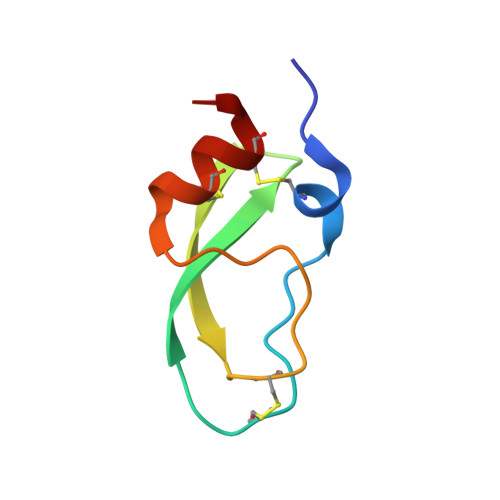 | |
UniProt | |||||
Entity Groups | |||||
| Sequence Clusters | 30% Identity50% Identity70% Identity90% Identity95% Identity100% Identity | ||||
| UniProt Group | P81658 | ||||
Sequence AnnotationsExpand | |||||
Reference Sequence | |||||
| Length ( Å ) | Angle ( ˚ ) |
|---|---|
| a = 95.991 | α = 90 |
| b = 95.991 | β = 90 |
| c = 39.486 | γ = 90 |
| Software Name | Purpose |
|---|---|
| PHENIX | refinement |
| HKL-3000 | data collection |
| HKL-3000 | data reduction |
| PHENIX | model building |
| PHENIX | phasing |


