5YU3

Structural basis for recognition of L-lysine, L-ornithine, and L-2,4-diamino butyric acid by lysine cyclodeaminase


Experimental Data Snapshot

  • Method: X-RAY DIFFRACTION
  • Resolution: 1.79 Å
  • R-Value Free: 0.193 
  • R-Value Work: 0.166 
  • R-Value Observed: 0.167 

wwPDB Validation   3D Report Full Report


Ligand Structure Quality Assessment 


This is version 1.2 of the entry. See complete history


Literature

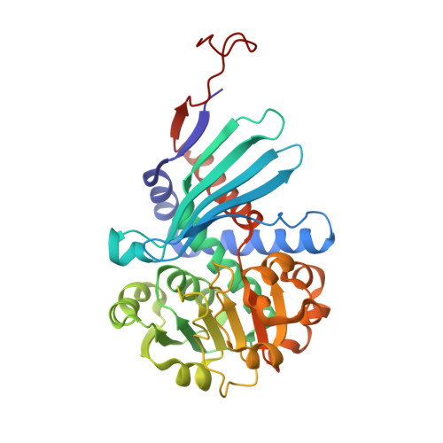
Structural Basis for Recognition of L-lysine, L-ornithine, and L-2,4-diamino Butyric Acid by Lysine Cyclodeaminase.

Min, K.Yoon, H.J.Matsuura, A.Kim, Y.H.Lee, H.H.

(2018) Mol Cells 41: 331-341

  • DOI: https://doi.org/10.14348/molcells.2018.2313
  • Primary Citation of Related Structures:  
    5YU0, 5YU1, 5YU3, 5YU4

  • PubMed Abstract: 

    L-pipecolic acid is a non-protein amino acid commonly found in plants, animals, and microorganisms. It is a well-known precursor to numerous microbial secondary metabolites and pharmaceuticals, including anticancer agents, immunosuppressants, and several antibiotics. Lysine cyclodeaminase (LCD) catalyzes β-deamination of L-lysine into L-pipecolic acid using β-nicotinamide adenine dinucleotide as a cofactor. Expression of a human homolog of LCD, μ-crystallin, is elevated in prostate cancer patients. To understand the structural features and catalytic mechanisms of LCD, we determined the crystal structures of Streptomyces pristinaespiralis LCD (SpLCD) in (i) a binary complex with NAD + , (ii) a ternary complex with NAD + and L-pipecolic acid, (iii) a ternary complex with NAD + and L-proline, and (iv) a ternary complex with NAD + and L-2,4-diamino butyric acid. The overall structure of SpLCD was similar to that of ornithine cyclodeaminase from Pseudomonas putida . In addition, SpLCD recognized L-lysine, L-ornithine, and L-2,4-diamino butyric acid despite differences in the active site, including differences in hydrogen bonding by Asp236, which corresponds with Asp228 from Pseudomonas putida ornithine cyclodeaminase. The substrate binding pocket of SpLCD allowed substrates smaller than lysine to bind, thus enabling binding to ornithine and L-2,4-diamino butyric acid. Our structural and biochemical data facilitate a detailed understanding of substrate and product recognition, thus providing evidence for a reaction mechanism for SpLCD. The proposed mechanism is unusual in that NAD + is initially converted into NADH and then reverted back into NAD + at a late stage of the reaction.


  • Organizational Affiliation

    Department of Chemistry, College of Natural Sciences, Seoul National University, Seoul 08826, Korea.


Macromolecules
Find similar proteins by:  (by identity cutoff)  |  3D Structure
Entity ID: 1
MoleculeChains Sequence LengthOrganismDetailsImage
Lysine cyclodeaminase
A, B, C, D
344Streptomyces pristinaespiralisMutation(s): 0 
Gene Names: pipASPRI_0308SPRI_7045
EC: 4.3.1.12
UniProt
Find proteins for D9UBW0 (Streptomyces pristinaespiralis)
Explore D9UBW0 
Go to UniProtKB:  D9UBW0
Entity Groups  
Sequence Clusters30% Identity50% Identity70% Identity90% Identity95% Identity100% Identity
UniProt GroupD9UBW0
Sequence Annotations
Expand
  • Reference Sequence
Small Molecules
Ligands 3 Unique
IDChains Name / Formula / InChI Key2D Diagram3D Interactions
NAD
Query on NAD

Download Ideal Coordinates CCD File 
F [auth A],
I [auth B],
L [auth C],
O [auth D]
NICOTINAMIDE-ADENINE-DINUCLEOTIDE
C21 H27 N7 O14 P2
BAWFJGJZGIEFAR-NNYOXOHSSA-N
PRO
Query on PRO

Download Ideal Coordinates CCD File 
G [auth A],
J [auth B],
M [auth C],
P [auth D]
PROLINE
C5 H9 N O2
ONIBWKKTOPOVIA-BYPYZUCNSA-N
NA
Query on NA

Download Ideal Coordinates CCD File 
E [auth A],
H [auth B],
K [auth C],
N [auth D]
SODIUM ION
Na
FKNQFGJONOIPTF-UHFFFAOYSA-N
Experimental Data & Validation

Experimental Data

  • Method: X-RAY DIFFRACTION
  • Resolution: 1.79 Å
  • R-Value Free: 0.193 
  • R-Value Work: 0.166 
  • R-Value Observed: 0.167 
  • Space Group: C 1 2 1
Unit Cell:
Length ( Å )Angle ( ˚ )
a = 271.529α = 90
b = 64.783β = 104.2
c = 106.858γ = 90
Software Package:
Software NamePurpose
PHENIXrefinement
HKL-2000data reduction
HKL-2000data scaling
PHENIXphasing

Structure Validation

View Full Validation Report


Ligand Structure Quality Assessment 


Entry History 

Deposition Data

Revision History  (Full details and data files)

  • Version 1.0: 2018-05-02
    Type: Initial release
  • Version 1.1: 2018-05-16
    Changes: Data collection, Database references
  • Version 1.2: 2024-03-27
    Changes: Data collection, Database references, Derived calculations