Manganese(V) Porphycene Complex Responsible for Inert C-H Bond Hydroxylation in a Myoglobin Matrix.
Oohora, K., Meichin, H., Kihira, Y., Sugimoto, H., Shiro, Y., Hayashi, T.(2017) J Am Chem Soc 139: 18460-18463
- PubMed: 29237270 Search on PubMed
- DOI: https://doi.org/10.1021/jacs.7b11288
- Primary Citation Related Structures:
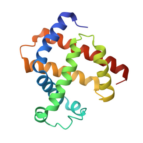
5YL3 - PubMed Abstract:
A mechanistic study of H 2 O 2 -dependent C-H bond hydroxylation by myoglobin reconstituted with a manganese porphycene was carried out. The X-ray crystal structure of the reconstituted protein obtained at 1.5 Å resolution reveals tight incorporation of the complex into the myoglobin matrix at pH 8.5, the optimized pH value for the highest turnover number of hydroxylation of ethylbenzene. The protein generates a spectroscopically detectable two-electron oxidative intermediate in a reaction with peracid, which has a half-life up to 38 s at 10 °C. Electron paramagnetic resonance spectra of the intermediate with perpendicular and parallel modes are silent, indicating formation of a low-spin Mn V -oxo species. In addition, the Mn V -oxo species is capable of promoting the hydroxylation of sodium 4-ethylbenzenesulfonate under single turnover conditions with an apparent second-order rate constant of 2.0 M -1 s -1 at 25 °C. Furthermore, the higher bond dissociation enthalpy of the substrate decreases the rate constant, in support of the proposal that the H-abstraction is one of the rate-limiting steps. The present engineered myoglobin serves as an artificial metalloenzyme for inert C-H bond activation via a high-valent metal species similar to the species employed by native monooxygenases such as cytochrome P450.
- Department of Applied Chemistry, Graduate School of Engineering, Osaka University , Suita 565-0871, Japan.
Organizational Affiliation: