5YI8

Crystal structure of drosophila Numb PTB domain and Pon peptide complex


Experimental Data Snapshot

  • Method: X-RAY DIFFRACTION
  • Resolution: 2.00 Å
  • R-Value Free: 0.227 
  • R-Value Work: 0.191 
  • R-Value Observed: 0.193 

wwPDB Validation   3D Report Full Report


Ligand Structure Quality Assessment 


This is version 1.1 of the entry. See complete history


Literature

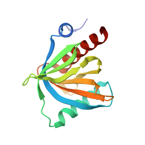
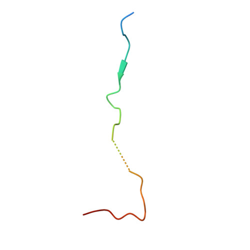
Basal condensation of Numb and Pon complex via phase transition during Drosophila neuroblast asymmetric division.

Shan, Z.Tu, Y.Yang, Y.Liu, Z.Zeng, M.Xu, H.Long, J.Zhang, M.Cai, Y.Wen, W.

(2018) Nat Commun 9: 737-737

  • DOI: https://doi.org/10.1038/s41467-018-03077-3
  • Primary Citation of Related Structures:  
    5YI7, 5YI8

  • PubMed Abstract: 

    Uneven distribution and local concentration of protein complexes on distinct membrane cortices is a fundamental property in numerous biological processes, including Drosophila neuroblast (NB) asymmetric cell divisions and cell polarity in general. In NBs, the cell fate determinant Numb forms a basal crescent together with Pon and is segregated into the basal daughter cell to initiate its differentiation. Here we discover that Numb PTB domain, using two distinct binding surfaces, recognizes repeating motifs within Pon in a previously unrecognized mode. The multivalent Numb-Pon interaction leads to high binding specificity and liquid-liquid phase separation of the complex. Perturbations of the Numb/Pon complex phase transition impair the basal localization of Numb and its subsequent suppression of Notch signaling during NB asymmetric divisions. Such phase-transition-mediated protein condensations on distinct membrane cortices may be a general mechanism for various cell polarity regulatory complexes.


  • Organizational Affiliation

    Department of Neurosurgery, Huashan Hospital, Institutes of Biomedical Sciences, State Key Laboratory of Medical Neurobiology, Department of Systems Biology for Medicine, School of Basic Medical Sciences, Shanghai Medical College of Fudan University, Shanghai, 200032, China.


Macromolecules
Find similar proteins by:  (by identity cutoff)  |  3D Structure
Entity ID: 1
MoleculeChains Sequence LengthOrganismDetailsImage
Protein numb141Drosophila melanogasterMutation(s): 0 
Gene Names: numbCG3779
UniProt
Find proteins for P16554 (Drosophila melanogaster)
Explore P16554 
Go to UniProtKB:  P16554
Entity Groups  
Sequence Clusters30% Identity50% Identity70% Identity90% Identity95% Identity100% Identity
UniProt GroupP16554
Sequence Annotations
Expand
  • Reference Sequence
Find similar proteins by:  (by identity cutoff)  |  3D Structure
Entity ID: 2
MoleculeChains Sequence LengthOrganismDetailsImage
Pon peptide from Partner of numb33Drosophila melanogasterMutation(s): 0 
UniProt
Find proteins for Q9W4I7 (Drosophila melanogaster)
Explore Q9W4I7 
Go to UniProtKB:  Q9W4I7
Entity Groups  
Sequence Clusters30% Identity50% Identity70% Identity90% Identity95% Identity100% Identity
UniProt GroupQ9W4I7
Sequence Annotations
Expand
  • Reference Sequence
Small Molecules
Ligands 1 Unique
IDChains Name / Formula / InChI Key2D Diagram3D Interactions
FMT (Subject of Investigation/LOI)
Query on FMT

Download Ideal Coordinates CCD File 
C [auth B]FORMIC ACID
C H2 O2
BDAGIHXWWSANSR-UHFFFAOYSA-N
Experimental Data & Validation

Experimental Data

  • Method: X-RAY DIFFRACTION
  • Resolution: 2.00 Å
  • R-Value Free: 0.227 
  • R-Value Work: 0.191 
  • R-Value Observed: 0.193 
  • Space Group: P 32 2 1
Unit Cell:
Length ( Å )Angle ( ˚ )
a = 43.392α = 90
b = 43.392β = 90
c = 181.304γ = 120
Software Package:
Software NamePurpose
PHENIXrefinement
HKL-2000data collection
HKL-2000data scaling
PHASERphasing
PDB_EXTRACTdata extraction

Structure Validation

View Full Validation Report


Ligand Structure Quality Assessment 


Entry History 

Deposition Data

Revision History  (Full details and data files)

  • Version 1.0: 2018-04-18
    Type: Initial release
  • Version 1.1: 2023-11-22
    Changes: Data collection, Database references, Refinement description