5Y6T

Crystal structure of endo-1,4-beta-mannanase from Eisenia fetida


Experimental Data Snapshot

  • Method: X-RAY DIFFRACTION
  • Resolution: 1.70 Å
  • R-Value Free: 0.169 
  • R-Value Work: 0.130 
  • R-Value Observed: 0.132 

wwPDB Validation   3D Report Full Report


This is version 1.2 of the entry. See complete history


Literature

Gene cloning, expression, and X-ray crystallographic analysis of a beta-mannanase from Eisenia fetida.

Ueda, M.Hirano, Y.Fukuhara, H.Naka, Y.Nakazawa, M.Sakamoto, T.Ogata, Y.Tamada, T.

(2018) Enzyme Microb Technol 117: 15-22

  • DOI: https://doi.org/10.1016/j.enzmictec.2018.05.014
  • Primary Citation of Related Structures:  
    5Y6T

  • PubMed Abstract: 

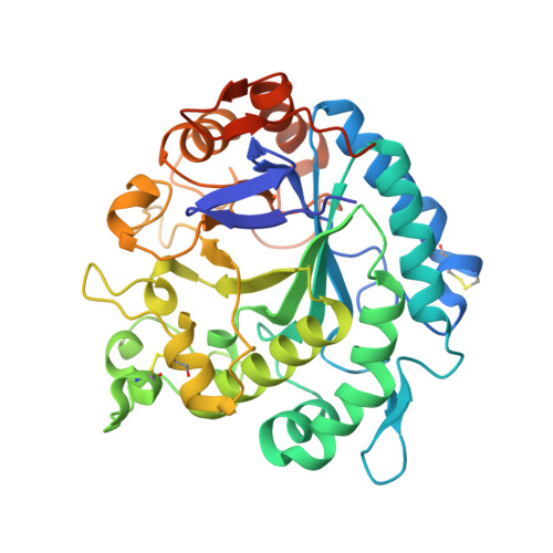
    The endo-1,4-β-mannanases (Ef-Man) gene from Eisenia fetida was determined to consist of 1131 bp and encode a 377 amino acid protein. The amino acid sequence showed similarity with the endo-1,4-β-mannanases of Daphnia pulex (62%), Cryptopygus antarcticus (64%), Crassostrea gigas (61%), Mytilus edulis (60%), and Aplysia kurodai (58%). The gene encoding mature Ef-Man was expressed in Pichia pastoris (GS115 strain). Based on SDS-PAGE analysis, the molecular mass of the purified recombinant Ef-Man (rEf-Man) was estimated to be 39 kDa. All catalytically important residues of endo-1,4-β-mannanases in the glycoside hydrolase (GH) family 5 were conserved in Ef-Man. The optimal temperature for rEf-Man was identified as 60 °C. HPLC and HPAEC analyses suggest that Ef-Man requires at least six subsites for efficient hydrolysis and is capable of performing transglycosylation reactions. The overall structure of rEf-Man is similar to those of GH5 family proteins, and tertiary structures around the active site are conserved among endo-1,4-β-mannanase families. X-ray crystallographic analysis supports the hydrolysis and transglycosylation reaction mechanism determined by HPLC and HPAEC analyses.


  • Organizational Affiliation

    Graduate School of Life and Environmental Sciences, Osaka Prefecture University, Osaka, 599-8531, Japan. Electronic address: mueda@biochem.osakafu-u.ac.jp.


Macromolecules
Find similar proteins by:  (by identity cutoff)  |  3D Structure
Entity ID: 1
MoleculeChains Sequence LengthOrganismDetailsImage
endo-1,4-beta-mannanase387Eisenia fetidaMutation(s): 0 
UniProt
Find proteins for A0A3B6UEQ6 (Eisenia fetida)
Explore A0A3B6UEQ6 
Go to UniProtKB:  A0A3B6UEQ6
Entity Groups  
Sequence Clusters30% Identity50% Identity70% Identity90% Identity95% Identity100% Identity
UniProt GroupA0A3B6UEQ6
Sequence Annotations
Expand
  • Reference Sequence
Small Molecules
Ligands 2 Unique
IDChains Name / Formula / InChI Key2D Diagram3D Interactions
TRS
Query on TRS

Download Ideal Coordinates CCD File 
B [auth A]2-AMINO-2-HYDROXYMETHYL-PROPANE-1,3-DIOL
C4 H12 N O3
LENZDBCJOHFCAS-UHFFFAOYSA-O
IPA
Query on IPA

Download Ideal Coordinates CCD File 
C [auth A]ISOPROPYL ALCOHOL
C3 H8 O
KFZMGEQAYNKOFK-UHFFFAOYSA-N
Experimental Data & Validation

Experimental Data

  • Method: X-RAY DIFFRACTION
  • Resolution: 1.70 Å
  • R-Value Free: 0.169 
  • R-Value Work: 0.130 
  • R-Value Observed: 0.132 
  • Space Group: P 21 21 21
Unit Cell:
Length ( Å )Angle ( ˚ )
a = 50.13α = 90
b = 69.461β = 90
c = 86.654γ = 90
Software Package:
Software NamePurpose
REFMACrefinement
XDSdata reduction
XDSdata scaling
PHASERphasing

Structure Validation

View Full Validation Report


Entry History & Funding Information

Deposition Data


Funding OrganizationLocationGrant Number
The Tojuro Iijima Memorial Foundation for Food Science and TechnologyJapan--
Japan Society for the Promotion of ScienceJapan--

Revision History  (Full details and data files)

  • Version 1.0: 2018-06-27
    Type: Initial release
  • Version 1.1: 2019-12-25
    Changes: Database references
  • Version 1.2: 2023-11-22
    Changes: Data collection, Database references, Refinement description