5XZF | pdb_00005xzf

Vitamin D receptor with a synthetic ligand ADRO1


Experimental Data Snapshot

  • Method: X-RAY DIFFRACTION
  • Resolution: 2.10 Å
  • R-Value Free: 
    0.214 (Depositor), 0.218 (DCC) 
  • R-Value Work: 
    0.161 (Depositor), 0.171 (DCC) 
  • R-Value Observed: 
    0.164 (Depositor) 

Starting Model: experimental
View more details

wwPDB Validation 3D Report Full Report

Validation slider image for 5XZF

Ligand Structure Quality Assessment 


This is version 1.3 of the entry. See complete history

Literature

25 S-Adamantyl-23-yne-26,27-dinor-1 alpha ,25-dihydroxyvitamin D3: Synthesis, Tissue Selective Biological Activities, and X-ray Crystal Structural Analysis of Its Vitamin D Receptor Complex.

Otero, R.Ishizawa, M.Numoto, N.Ikura, T.Ito, N.Tokiwa, H.Mourino, A.Makishima, M.Yamada, S.

(2018) J Med Chem 61: 6658-6673

  • DOI: https://doi.org/10.1021/acs.jmedchem.8b00427
  • Primary Citation Related Structures: 
    5XZF, 5XZH

  • PubMed Abstract: 

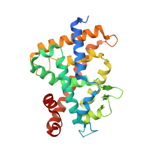
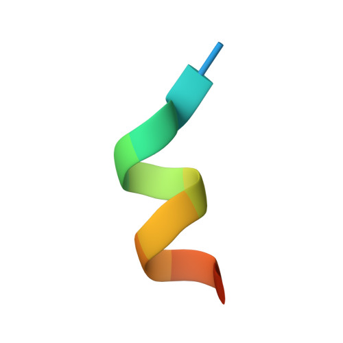
    Both 25 R- and 25 S-25-adamantyl-23-yne-26,27-dinor-1α,25-dihydroxyvitamin D 3 (4a and 4b) were stereoselectively synthesized by a Pd(0)-catalyzed ring closure and Suzuki-Miyaura coupling between enol-triflate 7 and alkenyl-boronic ester 8. The 25 S isomer (4b) showed high vitamin D receptor (VDR) affinity (50% of that of the natural hormone 1α,25-dihydroxyvitamin D 3 , 1) and transactivation potency (kidney HEK293, 90%). In endogenous gene expression, it showed high cell-type selectivity for kidney cells (HEK293, CYP24A1 160% of 1), bone cells (MG63, osteocalcin 64%), and monocytes (U937, CAMP 96%) over intestine (SW480, CYP24A1 8%) and skin (HaCaT, CYP24A1 7%) cells. The X-ray crystal structural analysis of 4b in complex with rat VDR-ligand binding domain (LBD) showed the highest Cα positional shift from the 1/VDR-LBD complex at helix 11. Helix 11 of the 4b and 1 VDR-LBD complexes also showed significant differences in surface properties. These results suggest that 4b should be examined further as another candidate for a mild preventive osteoporosis agent.


  • Organizational Affiliation
    • Departamento de Química Orgánica, Laboratorio de Investigación Ignacio Ribas , Universidad de Santiago de Compostela , 15782 Santiago de Compostela , Spain.

Macromolecule Content 

  • Total Structure Weight: 32.73 kDa 
  • Atom Count: 2,076 
  • Modeled Residue Count: 247 
  • Deposited Residue Count: 284 
  • Unique protein chains: 2

Macromolecules

Find similar proteins by:|  3D Structure
Entity ID: 1
MoleculeChains  Sequence LengthOrganismDetailsImage
Vitamin D3 receptor271Rattus norvegicusMutation(s): 0 
Gene Names: VdrNr1i1
UniProt
Find proteins for P13053 (Rattus norvegicus)
Explore P13053 
Go to UniProtKB:  P13053
Entity Groups
Sequence Clusters30% Identity50% Identity70% Identity90% Identity95% Identity100% Identity
UniProt GroupP13053
Sequence Annotations
Expand
Reference Sequence
Find similar proteins by:  Sequence   |   3D Structure  
Entity ID: 2
MoleculeChains  Sequence LengthOrganismDetailsImage
Mediator of RNA polymerase II transcription subunit 1B [auth C]13Homo sapiensMutation(s): 0 
UniProt & NIH Common Fund Data Resources
Find proteins for Q15648 (Homo sapiens)
Explore Q15648 
Go to UniProtKB:  Q15648
PHAROS:  Q15648
GTEx:  ENSG00000125686 
Entity Groups
Sequence Clusters30% Identity50% Identity70% Identity90% Identity95% Identity100% Identity
UniProt GroupQ15648
Sequence Annotations
Expand
Reference Sequence

Small Molecules

Ligands 2 Unique
IDChains Name / Formula / InChI Key2D Diagram3D Interactions
8J0

Query on 8J0


Download:Ideal Coordinates CCD File
C [auth A](1R,3S,5Z)-5-[(2E)-2-[(1R,3aS,7aR)-1-[(2R,6S)-6-(1-adamantyl)-6-oxidanyl-hex-4-yn-2-yl]-7a-methyl-2,3,3a,5,6,7-hexahydro-1H-inden-4-ylidene]ethylidene]-4-methylidene-cyclohexane-1,3-diol
C35 H50 O3
RKVGVUCXMUXZTG-LLLPRGGASA-N
FMT

Query on FMT


Download:Ideal Coordinates CCD File
D [auth A]FORMIC ACID
C H2 O2
BDAGIHXWWSANSR-UHFFFAOYSA-N

Experimental Data & Validation

Experimental Data

  • Method: X-RAY DIFFRACTION
  • Resolution: 2.10 Å
  • R-Value Free:  0.214 (Depositor), 0.218 (DCC) 
  • R-Value Work:  0.161 (Depositor), 0.171 (DCC) 
  • R-Value Observed: 0.164 (Depositor) 
Space Group: C 1 2 1
Unit Cell:
Length ( Å )Angle ( ˚ )
a = 154.07α = 90
b = 41.11β = 96.42
c = 41.42γ = 90
Software Package:
Software NamePurpose
REFMACrefinement
XDSdata reduction
XDSdata scaling
PHASERphasing

Structure Validation

View Full Validation Report


Ligand Structure Quality Assessment 


Entry History 

Deposition Data

Revision History  (Full details and data files)

  • Version 1.0: 2018-07-25
    Type: Initial release
  • Version 1.1: 2018-08-01
    Changes: Data collection, Database references
  • Version 1.2: 2018-08-29
    Changes: Data collection, Database references
  • Version 1.3: 2023-11-22
    Changes: Data collection, Database references, Refinement description