5XX1

Crystal structure of Arginine decarboxylase (AdiA) from Salmonella typhimurium


Experimental Data Snapshot

  • Method: X-RAY DIFFRACTION
  • Resolution: 3.10 Å
  • R-Value Free: 0.272 
  • R-Value Work: 0.241 
  • R-Value Observed: 0.242 

wwPDB Validation   3D Report Full Report


This is version 1.2 of the entry. See complete history


Literature

Structural studies on the decameric S. typhimurium arginine decarboxylase (ADC): Pyridoxal 5'-phosphate binding induces conformational changes

Deka, G.Bharath, S.R.Savithri, H.S.Murthy, M.R.N.

(2017) Biochem Biophys Res Commun 490: 1362-1368

  • DOI: https://doi.org/10.1016/j.bbrc.2017.07.032
  • Primary Citation of Related Structures:  
    5XX1, 6AA9

  • PubMed Abstract: 

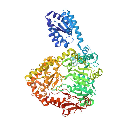
    Enteric pathogens such as Salmonella typhimurium colonize the human gut in spite of the lethal acidic pH environment (pH < 2.5) due to the activation of inducible acid tolerance response (ATR) systems. The pyridoxal 5'-phosphate (PLP)-dependent enzyme, biodegradative arginine decarboxylase (ADC, encoded by AdiA), is a component of an ATR system. The enzyme consumes a cytoplasmic proton in the process of arginine degradation to agmatine. Arginine-agmatine antiporter (AdiC) exchanges the product agmatine for arginine. In this manuscript, we describe the structure of Salmonella typhimurium ADC (StADC). The decameric structure assembled from five dimers related by a non crystallographic 5-fold symmetry represents the first apo-form of the enzyme. The structure suggests that PLP-binding is not a prerequisite for oligomerization. Comparison with E. coli ADC reveals that PLP-binding is accompanied by the movement and ordering of two loops (residues 150-159 and 191-197) and a few active site residues such as His256 and Lys257. A number of residues important for substrate binding are disordered in the apo-StADC structure indicating that PLP binding is important for substrate binding. Unlike the interactions between 5-fold related protomers, interactions that stabilize the dimeric structure are not pH dependent.


  • Organizational Affiliation

    Molecular Biophysics Unit, Indian Institute of Science, Bangalore 560012, India.


Macromolecules
Find similar proteins by:  (by identity cutoff)  |  3D Structure
Entity ID: 1
MoleculeChains Sequence LengthOrganismDetailsImage
Arginine decarboxylase
A, B, C, D, E
A, B, C, D, E, F, G, H, I, J
756Salmonella enterica subsp. enterica serovar TyphiMutation(s): 1 
Gene Names: adit4203
EC: 4.1.1.19
UniProt
Find proteins for Q8Z1P1 (Salmonella typhi)
Explore Q8Z1P1 
Go to UniProtKB:  Q8Z1P1
Entity Groups  
Sequence Clusters30% Identity50% Identity70% Identity90% Identity95% Identity100% Identity
UniProt GroupQ8Z1P1
Sequence Annotations
Expand
  • Reference Sequence
Small Molecules
Modified Residues  1 Unique
IDChains TypeFormula2D DiagramParent
CME
Query on CME
A, B, C, D, E
A, B, C, D, E, F, G, H, I, J
L-PEPTIDE LINKINGC5 H11 N O3 S2CYS
Experimental Data & Validation

Experimental Data

  • Method: X-RAY DIFFRACTION
  • Resolution: 3.10 Å
  • R-Value Free: 0.272 
  • R-Value Work: 0.241 
  • R-Value Observed: 0.242 
  • Space Group: P 21 21 21
Unit Cell:
Length ( Å )Angle ( ˚ )
a = 179.36α = 90
b = 193.25β = 90
c = 280.16γ = 90
Software Package:
Software NamePurpose
REFMACrefinement
iMOSFLMdata reduction
SCALAdata scaling
PHASERphasing

Structure Validation

View Full Validation Report


Entry History & Funding Information

Deposition Data


Funding OrganizationLocationGrant Number
Department of Biotechnology (DBT)IndiaDBT 0425

Revision History  (Full details and data files)

  • Version 1.0: 2018-05-16
    Type: Initial release
  • Version 1.1: 2018-05-30
    Changes: Data collection, Database references
  • Version 1.2: 2023-11-22
    Changes: Data collection, Database references, Refinement description