5XUP | pdb_00005xup

Crystal structure of TRF1 and TERB1


Experimental Data Snapshot

  • Method: X-RAY DIFFRACTION
  • Resolution: 2.10 Å
  • R-Value Free: 
    0.221 (Depositor), 0.224 (DCC) 
  • R-Value Work: 
    0.177 (Depositor), 0.179 (DCC) 
  • R-Value Observed: 
    0.179 (Depositor) 

Starting Model: experimental
View more details

wwPDB Validation 3D Report Full Report

Validation slider image for 5XUP

This is version 1.2 of the entry. See complete history

Literature

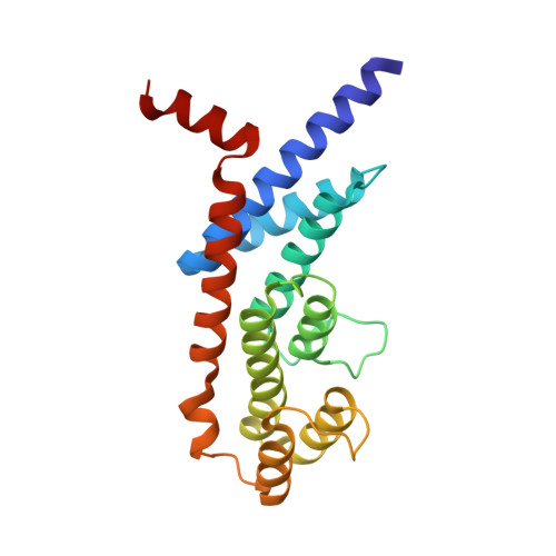
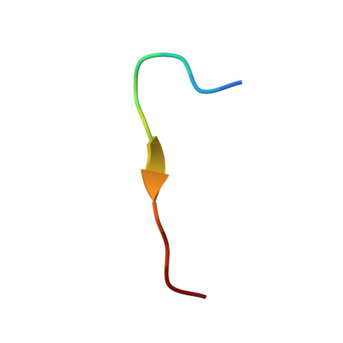
Telomeric TERB1-TRF1 interaction is crucial for male meiosis.

Long, J.Huang, C.Chen, Y.Zhang, Y.Shi, S.Wu, L.Liu, Y.Liu, C.Wu, J.Lei, M.

(2017) Nat Struct Mol Biol 24: 1073-1080

  • DOI: https://doi.org/10.1038/nsmb.3496
  • Primary Citation Related Structures: 
    5XUP

  • PubMed Abstract: 

    During meiotic prophase, the meiosis-specific telomere-binding protein TERB1 regulates chromosome movement required for homologous pairing and recombination by interacting with the telomeric shelterin subunit TRF1. Here, we report the crystal structure of the TRF1-binding motif of human TERB1 in complex with the TRFH domain of TRF1. Notably, specific disruption of the TERB1-TRF1 interaction by a point mutation in the mouse Terb1 gene results in infertility only in males. We find that this mutation causes an arrest in the zygotene-early pachytene stage and mild telomere abnormalities of autosomes but unpaired X and Y chromosomes in pachytene, leading to massive spermatocyte apoptosis. We propose that the loss of telomere structure mediated by the TERB1-TRF1 interaction significantly affects homologous pairing of the telomere-adjacent pseudoautosomal region (PAR) of the X and Y chromosomes in mouse spermatocytes. Our findings uncover a specific mechanism of telomeres that surmounts the unique challenges of mammalian X-Y pairing in meiosis.


  • Organizational Affiliation
    • National Center for Protein Science Shanghai, State Key Laboratory of Molecular Biology, Institute of Biochemistry and Cell Biology, Shanghai Institutes for Biological Sciences, Chinese Academy of Sciences, Shanghai, China.

Macromolecule Content 

  • Total Structure Weight: 49.31 kDa 
  • Atom Count: 3,509 
  • Modeled Residue Count: 416 
  • Deposited Residue Count: 428 
  • Unique protein chains: 2

Macromolecules

Find similar proteins by:|  3D Structure
Entity ID: 1
MoleculeChains  Sequence LengthOrganismDetailsImage
Telomeric repeat-binding factor 1
A, B
202Homo sapiensMutation(s): 0 
Gene Names: TERF1PIN2TRBF1TRFTRF1
UniProt & NIH Common Fund Data Resources
Find proteins for P54274 (Homo sapiens)
Explore P54274 
Go to UniProtKB:  P54274
PHAROS:  P54274
GTEx:  ENSG00000147601 
Entity Groups
Sequence Clusters30% Identity50% Identity70% Identity90% Identity95% Identity100% Identity
UniProt GroupP54274
Sequence Annotations
Expand
Reference Sequence
Find similar proteins by:  Sequence   |   3D Structure  
Entity ID: 2
MoleculeChains  Sequence LengthOrganismDetailsImage
Telomere repeats-binding bouquet formation protein 1
C, D
12Homo sapiensMutation(s): 0 
Gene Names: TERB1CCDC79
UniProt & NIH Common Fund Data Resources
Find proteins for Q8NA31 (Homo sapiens)
Explore Q8NA31 
Go to UniProtKB:  Q8NA31
PHAROS:  Q8NA31
GTEx:  ENSG00000249961 
Entity Groups
Sequence Clusters30% Identity50% Identity70% Identity90% Identity95% Identity100% Identity
UniProt GroupQ8NA31
Sequence Annotations
Expand
Reference Sequence

Experimental Data & Validation

Experimental Data

  • Method: X-RAY DIFFRACTION
  • Resolution: 2.10 Å
  • R-Value Free:  0.221 (Depositor), 0.224 (DCC) 
  • R-Value Work:  0.177 (Depositor), 0.179 (DCC) 
  • R-Value Observed: 0.179 (Depositor) 
Space Group: P 64
Unit Cell:
Length ( Å )Angle ( ˚ )
a = 161.579α = 90
b = 161.579β = 90
c = 45.925γ = 120
Software Package:
Software NamePurpose
HKL-2000data scaling
REFMACrefinement
PDB_EXTRACTdata extraction
HKL-2000data collection
MLPHAREmodel building

Structure Validation

View Full Validation Report


Entry History 

Deposition Data

Revision History  (Full details and data files)

  • Version 1.0: 2017-11-01
    Type: Initial release
  • Version 1.1: 2017-12-20
    Changes: Database references
  • Version 1.2: 2023-11-22
    Changes: Data collection, Database references, Refinement description