5XSW

Crystal structure of an archaeal chitinase in the substrate-complex form (P63)


Experimental Data Snapshot

  • Method: X-RAY DIFFRACTION
  • Resolution: 1.95 Å
  • R-Value Free: 0.196 
  • R-Value Work: 0.167 
  • R-Value Observed: 0.168 

wwPDB Validation   3D Report Full Report


This is version 2.1 of the entry. See complete history


Literature

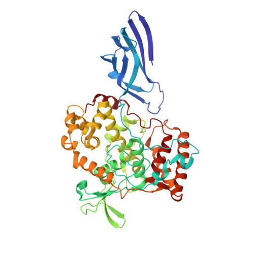
Crystal structures of an archaeal chitinase ChiD and its ligand complexes.

Nishitani, Y.Horiuchi, A.Aslam, M.Kanai, T.Atomi, H.Miki, K.

(2018) Glycobiology 28: 418-426

  • DOI: https://doi.org/10.1093/glycob/cwy024
  • Primary Citation of Related Structures:  
    5XSV, 5XSW, 5XSX

  • PubMed Abstract: 

    Chitinase D (designated as Pc-ChiD) was found in a hyperthermophilic archaeon, Pyrococcus chitonophagus (previously described as Thermococcus chitonophagus), that was isolated from media containing only chitin as carbon source. Pc-ChiD displays chitinase activity and is thermostable at temperatures up to 95°C, suggesting its potential for industrial use. Pc-ChiD has a secretion signal peptide and two chitin-binding domains (ChBDs) in the N-terminal domain. However, the C-terminal domain shares no sequence similarity with previously identified saccharide-degrading enzymes and does not contain the DXDXE motif conserved in the glycoside hydrolase (GH) 18 family chitinases. To elucidate its overall structure and reaction mechanism, we determined the first crystal structures of Pc-ChiD, both in the ligand-free form and in complexes with substrates. Structure analyses revealed that the C-terminal domain of Pc-ChiD, Pc-ChiD(ΔBD), consists of a third putative substrate-binding domain, which cannot be predicted from the amino acid sequence, and a catalytic domain structurally similar to that found in not the GH18 family but the GH23 family. Based on the similarity with GH23 family chitinase, the catalytic residues of Pc-ChiD were predicted and confirmed by mutagenesis analyses. Moreover, the specific C-terminal 100 residues of Pc-ChiD are important to fix the putative substrate-binding domain next to the catalytic domain, contributing to the structure stability as well as the long chitin chain binding. Our findings reveal the structure of a unique archaeal chitinase that is distinct from previously known members of the GH23 family.


  • Organizational Affiliation

    Department of Chemistry, Graduate School of Science, Kyoto University, Sakyo-ku, Kyoto 606-8502, Japan.


Macromolecules
Find similar proteins by:  (by identity cutoff)  |  3D Structure
Entity ID: 1
MoleculeChains Sequence LengthOrganismDetailsImage
Chitinase
A, B
486Thermococcus chitonophagusMutation(s): 0 
Gene Names: chiDCHITON_1119
EC: 3.2.1.14
UniProt
Find proteins for A0A161KIT4 (Thermococcus chitonophagus)
Explore A0A161KIT4 
Go to UniProtKB:  A0A161KIT4
Entity Groups  
Sequence Clusters30% Identity50% Identity70% Identity90% Identity95% Identity100% Identity
UniProt GroupA0A161KIT4
Sequence Annotations
Expand
  • Reference Sequence
Oligosaccharides

Help

Entity ID: 2
MoleculeChains Length2D Diagram Glycosylation3D Interactions
2-acetamido-2-deoxy-beta-D-glucopyranose-(1-4)-2-acetamido-2-deoxy-alpha-D-glucopyranose
C, D
2N/A
Glycosylation Resources
GlyTouCan:  G05026ZL
GlyCosmos:  G05026ZL
GlyGen:  G05026ZL
Small Molecules
Ligands 2 Unique
IDChains Name / Formula / InChI Key2D Diagram3D Interactions
PO4
Query on PO4

Download Ideal Coordinates CCD File 
E [auth A],
F [auth A],
M [auth B],
N [auth B]
PHOSPHATE ION
O4 P
NBIIXXVUZAFLBC-UHFFFAOYSA-K
GOL
Query on GOL

Download Ideal Coordinates CCD File 
G [auth A]
H [auth A]
I [auth A]
J [auth A]
K [auth A]
G [auth A],
H [auth A],
I [auth A],
J [auth A],
K [auth A],
L [auth A],
O [auth B],
P [auth B],
Q [auth B],
R [auth B],
S [auth B],
T [auth B]
GLYCEROL
C3 H8 O3
PEDCQBHIVMGVHV-UHFFFAOYSA-N
Experimental Data & Validation

Experimental Data

  • Method: X-RAY DIFFRACTION
  • Resolution: 1.95 Å
  • R-Value Free: 0.196 
  • R-Value Work: 0.167 
  • R-Value Observed: 0.168 
  • Space Group: P 63
Unit Cell:
Length ( Å )Angle ( ˚ )
a = 182.253α = 90
b = 182.253β = 90
c = 110.791γ = 120
Software Package:
Software NamePurpose
HKL-2000data processing
SOLVEphasing
RESOLVEphasing
Cootmodel building
PHENIXrefinement

Structure Validation

View Full Validation Report


Entry History & Funding Information

Deposition Data


Funding OrganizationLocationGrant Number
CRESTJapan--

Revision History  (Full details and data files)

  • Version 1.0: 2018-05-02
    Type: Initial release
  • Version 1.1: 2018-06-13
    Changes: Data collection, Database references
  • Version 2.0: 2020-07-29
    Type: Remediation
    Reason: Carbohydrate remediation
    Changes: Atomic model, Data collection, Derived calculations, Structure summary
  • Version 2.1: 2024-03-27
    Changes: Data collection, Database references, Derived calculations, Structure summary