5XPN | pdb_00005xpn

Crystal structure of VDR-LBD complexed with 25RS-(hydroxyphenyl)-25-methoxy-2-methylidene-19,26,27-trinor-1-hydroxyvitamin D3


Experimental Data Snapshot

  • Method: X-RAY DIFFRACTION
  • Resolution: 1.96 Å
  • R-Value Free: 
    0.256 (Depositor), 0.265 (DCC) 
  • R-Value Work: 
    0.207 (Depositor), 0.220 (DCC) 
  • R-Value Observed: 
    0.210 (Depositor) 

wwPDB Validation 3D Report Full Report

Validation slider image for 5XPN

Ligand Structure Quality Assessment 


This is version 1.1 of the entry. See complete history

Literature

Vitamin D Analogues with a p-Hydroxyphenyl Group at the C25 Position: Crystal Structure of Vitamin D Receptor Ligand-Binding Domain Complexed with the Ligand Explains the Mechanism Underlying Full Antagonistic Action

Kato, A.Yamao, M.Hashihara, Y.Ishida, H.Itoh, T.Yamamoto, K.

(2017) J Med Chem 60: 8394-8406

  • DOI: https://doi.org/10.1021/acs.jmedchem.7b00819
  • Primary Citation Related Structures: 
    5XPL, 5XPM, 5XPN, 5XPO, 5XPP

  • PubMed Abstract: 

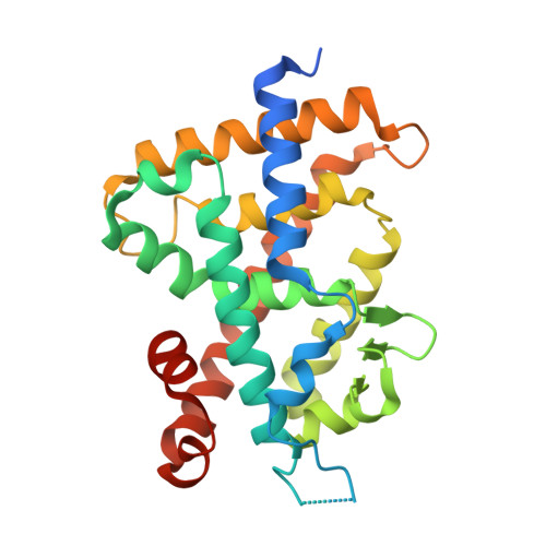
    Vitamin D receptor (VDR) antagonists can be classified into two categories: the first category of VDR antagonists, which do not stabilize the helix 11-12, and the second category of antagonists, which destabilize the helix 6-7 region. To elucidate the mechanism underlying the first category antagonists by using the crystal structure, we designed and synthesized several VDR ligands with a p-hydroxyphenyl group at the C25-position. Of these, 22S-butyl-25-carbonyl analogue 5b and 25-di-p-hydoroxyphenyl analogues 6a,b showed strong antagonistic activity. We succeeded in cocrystallizing the ligand-binding domain of VDR complexed with 5b and found that the structure showed an alternative conformation of the helix 11-12 that explained the mechanism of the first category antagonists. Taking the present and previous studies together, we could elucidate the mechanisms underlying first and second categories antagonists based on individual crystal structures. This study provides significant insights into antagonism against not only VDR but also nuclear receptors.


  • Organizational Affiliation
    • Laboratory of Drug Design and Medicinal Chemistry, Showa Pharmaceutical University , 3-3165 Higashi-Tamagawagakuen, Machida, Tokyo 194-8543, Japan.

Macromolecule Content 

  • Total Structure Weight: 33.16 kDa 
  • Atom Count: 2,231 
  • Modeled Residue Count: 250 
  • Deposited Residue Count: 284 
  • Unique protein chains: 2

Macromolecules

Find similar proteins by:|  3D Structure
Entity ID: 1
MoleculeChains  Sequence LengthOrganismDetailsImage
Vitamin D3 receptor271Rattus norvegicusMutation(s): 0 
Gene Names: VdrNr1i1
UniProt
Find proteins for P13053 (Rattus norvegicus)
Explore P13053 
Go to UniProtKB:  P13053
Entity Groups
Sequence Clusters30% Identity50% Identity70% Identity90% Identity95% Identity100% Identity
UniProt GroupP13053
Sequence Annotations
Expand
Reference Sequence
Find similar proteins by:  Sequence   |   3D Structure  
Entity ID: 2
MoleculeChains  Sequence LengthOrganismDetailsImage
Mediator of RNA polymerase II transcription subunit 1B [auth C]13Homo sapiensMutation(s): 0 
Gene Names: 
UniProt & NIH Common Fund Data Resources
Find proteins for Q15648 (Homo sapiens)
Explore Q15648 
Go to UniProtKB:  Q15648
PHAROS:  Q15648
GTEx:  ENSG00000125686 
Entity Groups
Sequence Clusters30% Identity50% Identity70% Identity90% Identity95% Identity100% Identity
UniProt GroupQ15648
Sequence Annotations
Expand
Reference Sequence

Small Molecules

Ligands 2 Unique
IDChains Name / Formula / InChI Key2D Diagram3D Interactions
9RO

Query on 9RO


Download:Ideal Coordinates CCD File
D [auth A](1~{R},3~{R})-5-[(2~{E})-2-[(1~{R},3~{a}~{S},7~{a}~{R})-1-[(2~{R},6~{R})-6-(4-hydroxyphenyl)-6-methoxy-hexan-2-yl]-7~{a}-methyl-2,3,3~{a},5,6,7-hexahydro-1~{H}-inden-4-ylidene]ethylidene]-2-methylidene-cyclohexane-1,3-diol
C32 H46 O4
LNLTYAYGVNOEBQ-OJABYWBNSA-N
8BO

Query on 8BO


Download:Ideal Coordinates CCD File
C [auth A](1~{R},3~{R})-5-[(2~{E})-2-[(1~{R},3~{a}~{S},7~{a}~{R})-1-[(2~{R},6~{S})-6-(4-hydroxyphenyl)-6-methoxy-hexan-2-yl]-7~{a}-methyl-2,3,3~{a},5,6,7-hexahydro-1~{H}-inden-4-ylidene]ethylidene]-2-methylidene-cyclohexane-1,3-diol
C32 H46 O4
LNLTYAYGVNOEBQ-CLQLOTFESA-N

Experimental Data & Validation

Experimental Data

  • Method: X-RAY DIFFRACTION
  • Resolution: 1.96 Å
  • R-Value Free:  0.256 (Depositor), 0.265 (DCC) 
  • R-Value Work:  0.207 (Depositor), 0.220 (DCC) 
  • R-Value Observed: 0.210 (Depositor) 
Space Group: C 1 2 1
Unit Cell:
Length ( Å )Angle ( ˚ )
a = 153.74α = 90
b = 43.16β = 95.32
c = 42.03γ = 90
Software Package:
Software NamePurpose
REFMACrefinement

Structure Validation

View Full Validation Report


Ligand Structure Quality Assessment 


Entry History 

Deposition Data

Revision History  (Full details and data files)

  • Version 1.0: 2018-07-11
    Type: Initial release
  • Version 1.1: 2024-03-27
    Changes: Data collection, Database references