Structural modulation of a periplasmic sugar-binding protein probes into its evolutionary ancestry.
Pandey, S., Phale, P.S., Bhaumik, P.(2018) J Struct Biol 204: 498-506
- PubMed: 30244006 Search on PubMed
- DOI: https://doi.org/10.1016/j.jsb.2018.09.006
- Primary Citation Related Structures:
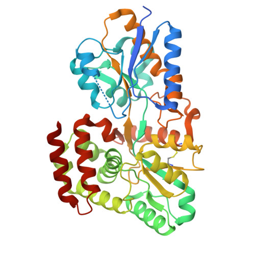
5XPJ - PubMed Abstract:
Substrate-binding proteins (SBPs) are periplasmic proteins consisting of two α/β domains joined by a hinge region with specificity towards cognate ligands. Based on three-dimensional fold, sugar-specific SBPs have been classified into cluster B and cluster D-I. The analysis of sequences and structures of sugar-binding pocket of cluster D-I SBPs revealed the presence of extra residues on two loops (L1, L2) and a helix (H1) in few members of this family, that binds specifically to monosaccharides. Presence of conserved histidine in L2 and tryptophan in H1 can be considered as the identity marks for the cluster D-I monosaccharide-binding SBPs. A glucose binding protein (ppGBP) from Pseudomonas putida CSV86 was found to contain a structural fold similar to oligosaccharide-binding cluster D-I SBPs, but functionally binds to only glucose due to constriction of its binding pocket mainly by L2 (375-382). ppGBP with partial deletion of L2 (ppGBPΔL2) was created, crystallized and biochemical characterization was performed. Compared to wild type ppGBP, the ppGBPΔL2 structure showed widening of the glucose-binding pocket with ∼80% lower glucose binding. Our results show that the substrate specificity of SBPs can be altered by modulating the size of the binding pocket. Based on this, we propose a sub classification of cluster D-I SBPs into (i) cluster D-I(a)-monosaccharide-binding SBPs and (ii) cluster D-I(b)-oligosaccharide-binding SBPs. This study also provides the direct structural and functional correlation indicating that divergence of proteins may occur through insertions or deletions of sequences in the already existing SBPs leading to evolution at the functional level.
- Department of Biosciences and Bioengineering, Indian Institute of Technology Bombay, Powai, Mumbai, Maharashtra 400076, India.
Organizational Affiliation: