5XM7 | pdb_00005xm7

Crystal structure of Plasmodium falciparum aminopeptidase N in complex with (S)-2-((2S,3R)-3-amino-2-hydroxy-4-phenylbutanamido)-N-hydroxy-4-methylpentanamide


Experimental Data Snapshot

  • Method: X-RAY DIFFRACTION
  • Resolution: 1.96 Å
  • R-Value Free: 
    0.219 (Depositor), 0.226 (DCC) 
  • R-Value Work: 
    0.168 (Depositor), 0.178 (DCC) 
  • R-Value Observed: 
    0.171 (Depositor) 

Starting Model: experimental
View more details

wwPDB Validation 3D Report Full Report

Validation slider image for 5XM7

Ligand Structure Quality Assessment 


This is version 1.2 of the entry. See complete history

Literature

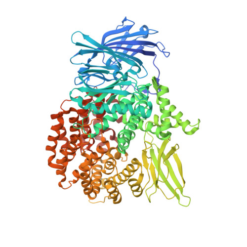
Development of peptidomimetic hydroxamates as PfA-M1 and PfA-M17 dual inhibitors: Biological evaluation and structural characterization by cocrystallization

Marapaka, A.K.Sankoju, P.Zhang, G.Ding, Y.Ma, C.Pillalamarri, V.Sudhakar, P.Reddi, B.Sijwali, P.S.Zhang, Y.Addlagatta, A.

(2022) Chin Chem Lett 33: 2550-2554

Macromolecule Content 

  • Total Structure Weight: 107.86 kDa 
  • Atom Count: 7,825 
  • Modeled Residue Count: 890 
  • Deposited Residue Count: 914 
  • Unique protein chains: 1

Macromolecules

Find similar proteins by:|  3D Structure
Entity ID: 1
MoleculeChains  Sequence LengthOrganismDetailsImage
M1 family aminopeptidase914Plasmodium falciparum FcB1/ColumbiaMutation(s): 0 
EC: 3.4.11
UniProt
Find proteins for O96935 (Plasmodium falciparum (isolate 3D7))
Explore O96935 
Go to UniProtKB:  O96935
Entity Groups
Sequence Clusters30% Identity50% Identity70% Identity90% Identity95% Identity100% Identity
UniProt GroupO96935
Sequence Annotations
Expand
Reference Sequence

Small Molecules

Ligands 4 Unique
IDChains Name / Formula / InChI Key2D Diagram3D Interactions
89X

Query on 89X


Download:Ideal Coordinates CCD File
Q [auth A](2S)-4-methyl-N-[(1R)-2-(oxidanylamino)-2-oxidanylidene-1-phenyl-ethyl]-2-[(phenylmethyl)carbamoylamino]pentanamide
C22 H28 N4 O4
HNVHIABPJCVMFJ-RBUKOAKNSA-N
GOL

Query on GOL


Download:Ideal Coordinates CCD File
F [auth A]
G [auth A]
H [auth A]
I [auth A]
J [auth A]
F [auth A],
G [auth A],
H [auth A],
I [auth A],
J [auth A],
K [auth A],
L [auth A],
M [auth A],
N [auth A],
O [auth A],
P [auth A]
GLYCEROL
C3 H8 O3
PEDCQBHIVMGVHV-UHFFFAOYSA-N
ZN

Query on ZN


Download:Ideal Coordinates CCD File
B [auth A]ZINC ION
Zn
PTFCDOFLOPIGGS-UHFFFAOYSA-N
MG

Query on MG


Download:Ideal Coordinates CCD File
C [auth A],
D [auth A],
E [auth A]
MAGNESIUM ION
Mg
JLVVSXFLKOJNIY-UHFFFAOYSA-N

Experimental Data & Validation

Experimental Data

  • Method: X-RAY DIFFRACTION
  • Resolution: 1.96 Å
  • R-Value Free:  0.219 (Depositor), 0.226 (DCC) 
  • R-Value Work:  0.168 (Depositor), 0.178 (DCC) 
  • R-Value Observed: 0.171 (Depositor) 
Space Group: P 21 21 21
Unit Cell:
Length ( Å )Angle ( ˚ )
a = 74.462α = 90
b = 109.103β = 90
c = 112.372γ = 90
Software Package:
Software NamePurpose
REFMACrefinement
DENZOdata collection
SCALEPACKdata scaling
MOLREPmodel building
PDB_EXTRACTdata extraction
DENZOdata reduction

Structure Validation

View Full Validation Report


Ligand Structure Quality Assessment 


Entry History 

Deposition Data

Revision History  (Full details and data files)

  • Version 1.0: 2018-06-20
    Type: Initial release
  • Version 1.1: 2022-07-27
    Changes: Database references, Derived calculations, Refinement description
  • Version 1.2: 2023-11-22
    Changes: Data collection, Refinement description