5XLW

Mycobacterium tuberculosis Pantothenate kinase mutant F247A/F254A


Experimental Data Snapshot

  • Method: X-RAY DIFFRACTION
  • Resolution: 2.26 Å
  • R-Value Free: 0.228 
  • R-Value Work: 0.191 
  • R-Value Observed: 0.193 

wwPDB Validation   3D Report Full Report


This is version 1.2 of the entry. See complete history


Literature

Biochemical and structural studies of mutants indicate concerted movement of the dimer interface and ligand-binding region of Mycobacterium tuberculosis pantothenate kinase

Paul, A.Kumar, P.Surolia, A.Vijayan, M.

(2017) Acta Crystallogr F Struct Biol Commun 73: 635-643

  • DOI: https://doi.org/10.1107/S2053230X17015667
  • Primary Citation of Related Structures:  
    5XLV, 5XLW, 5XMB

  • PubMed Abstract: 

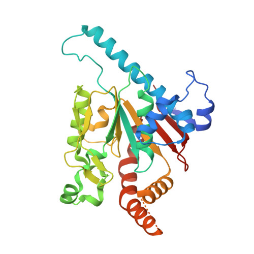
    Two point mutants and the corresponding double mutant of Mycobacterium tuberculosis pantothenate kinase have been prepared and biochemically and structurally characterized. The mutants were designed to weaken the affinity of the enzyme for the feedback inhibitor CoA. The mutants exhibit reduced activity, which can be explained in terms of their structures. The crystals of the mutants are not isomorphous to any of the previously analysed crystals of the wild-type enzyme or its complexes. The mycobacterial enzyme and its homologous Escherichia coli enzyme exhibit structural differences in their nucleotide complexes in the dimer interface and the ligand-binding region. In three of the four crystallographically independent mutant molecules the structure is similar to that in the E. coli enzyme. Although the mutants involve changes in the CoA-binding region, the dimer interface and the ligand-binding region move in a concerted manner, an observation which might be important in enzyme action. This work demonstrates that the structure of the mycobacterial enzyme can be transformed into a structure similar to that of the E. coli enzyme through minor perturbations without external influences such as those involving ligand binding.


  • Organizational Affiliation

    Molecular Biophysics Unit, Indian Institute of Science, Bangalore 560 012, India.


Macromolecules
Find similar proteins by:  (by identity cutoff)  |  3D Structure
Entity ID: 1
MoleculeChains Sequence LengthOrganismDetailsImage
Pantothenate kinaseA [auth B],
B [auth A]
312Mycobacterium tuberculosis H37RvMutation(s): 2 
Gene Names: coaARv1092cMTV017.45c
EC: 2.7.1.33
UniProt
Find proteins for P9WPA7 (Mycobacterium tuberculosis (strain ATCC 25618 / H37Rv))
Explore P9WPA7 
Go to UniProtKB:  P9WPA7
Entity Groups  
Sequence Clusters30% Identity50% Identity70% Identity90% Identity95% Identity100% Identity
UniProt GroupP9WPA7
Sequence Annotations
Expand
  • Reference Sequence
Small Molecules
Ligands 4 Unique
IDChains Name / Formula / InChI Key2D Diagram3D Interactions
1PE
Query on 1PE

Download Ideal Coordinates CCD File 
O [auth B]PENTAETHYLENE GLYCOL
C10 H22 O6
JLFNLZLINWHATN-UHFFFAOYSA-N
PEG
Query on PEG

Download Ideal Coordinates CCD File 
R [auth A]DI(HYDROXYETHYL)ETHER
C4 H10 O3
MTHSVFCYNBDYFN-UHFFFAOYSA-N
SO4
Query on SO4

Download Ideal Coordinates CCD File 
AA [auth A],
BA [auth A],
P [auth B],
Q [auth B]
SULFATE ION
O4 S
QAOWNCQODCNURD-UHFFFAOYSA-L
EDO
Query on EDO

Download Ideal Coordinates CCD File 
C [auth B]
D [auth B]
E [auth B]
F [auth B]
G [auth B]
C [auth B],
D [auth B],
E [auth B],
F [auth B],
G [auth B],
H [auth B],
I [auth B],
J [auth B],
K [auth B],
L [auth B],
M [auth B],
N [auth B],
S [auth A],
T [auth A],
U [auth A],
V [auth A],
W [auth A],
X [auth A],
Y [auth A],
Z [auth A]
1,2-ETHANEDIOL
C2 H6 O2
LYCAIKOWRPUZTN-UHFFFAOYSA-N
Experimental Data & Validation

Experimental Data

  • Method: X-RAY DIFFRACTION
  • Resolution: 2.26 Å
  • R-Value Free: 0.228 
  • R-Value Work: 0.191 
  • R-Value Observed: 0.193 
  • Space Group: P 31 2 1
Unit Cell:
Length ( Å )Angle ( ˚ )
a = 119.33α = 90
b = 119.33β = 90
c = 127.44γ = 120
Software Package:
Software NamePurpose
REFMACrefinement
iMOSFLMdata reduction
SCALAdata scaling
PHASERphasing

Structure Validation

View Full Validation Report


Entry History 

Deposition Data

Revision History  (Full details and data files)

  • Version 1.0: 2018-05-16
    Type: Initial release
  • Version 1.1: 2018-12-05
    Changes: Data collection, Database references
  • Version 1.2: 2023-11-22
    Changes: Data collection, Database references, Refinement description