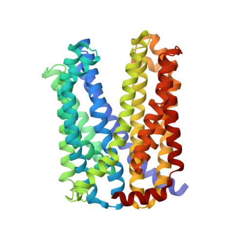
5XJJ

Crystal structure of a MATE family protein


Experimental Data Snapshot

  • Method: X-RAY DIFFRACTION
  • Resolution: 2.90 Å
  • R-Value Free: 0.296 
  • R-Value Work: 0.239 
  • R-Value Observed: 0.244 

wwPDB Validation   3D Report Full Report


Ligand Structure Quality Assessment 


This is version 1.1 of the entry. See complete history


Literature

Crystal Structure of a Plant Multidrug and Toxic Compound Extrusion Family Protein

Tanaka, Y.Iwaki, S.Tsukazaki, T.

(2017) Structure 25: 1455-1460.e2

  • DOI: 10.1016/j.str.2017.07.009
  • Primary Citation of Related Structures:  
    5XJJ, 5YCK

  • PubMed Abstract: 
  • The multidrug and toxic compound extrusion (MATE) family of proteins consists of transporters responsible for multidrug resistance in prokaryotes. In plants, a number of MATE proteins were identified by recent genomic and functional studies, which imply that the proteins have substrate-specific transport functions instead of multidrug extrusion ...

    The multidrug and toxic compound extrusion (MATE) family of proteins consists of transporters responsible for multidrug resistance in prokaryotes. In plants, a number of MATE proteins were identified by recent genomic and functional studies, which imply that the proteins have substrate-specific transport functions instead of multidrug extrusion. The three-dimensional structure of eukaryotic MATE proteins, including those of plants, has not been reported, preventing a better understanding of the molecular mechanism of these proteins. Here, we describe the crystal structure of a MATE protein from the plant Camelina sativa at 2.9 Å resolution. Two sets of six transmembrane α helices, assembled pseudo-symmetrically, possess a negatively charged internal pocket with an outward-facing shape. The crystal structure provides insight into the diversity of plant MATE proteins and their substrate recognition and transport through the membrane.


    Organizational Affiliation

    Graduate School of Biological Sciences, Nara Institute of Science and Technology, 8916-5 Takayama-cho, Ikoma, Nara 630-0192, Japan. Electronic address: ttsukazaki@mac.com.


Macromolecules
Find similar proteins by:  (by identity cutoff)  |  3D Structure
Entity ID: 1
MoleculeChainsSequence LengthOrganismDetailsImage
multi drug efflux transporterA455Camelina sativaMutation(s): 0 
Membrane Entity: Yes 
UniProt
Find proteins for A0A2D0TCI5 (Camelina sativa)
Explore A0A2D0TCI5 
Go to UniProtKB:  A0A2D0TCI5
Entity Groups  
Sequence Clusters30% Identity50% Identity70% Identity90% Identity95% Identity100% Identity
UniProt GroupA0A2D0TCI5
Protein Feature View
Expand
  • Reference Sequence
Small Molecules
Ligands 1 Unique
IDChainsName / Formula / InChI Key2D Diagram3D Interactions
OLC
Query on OLC

Download Ideal Coordinates CCD File 
B [auth A](2R)-2,3-dihydroxypropyl (9Z)-octadec-9-enoate
C21 H40 O4
RZRNAYUHWVFMIP-GDCKJWNLSA-N
 Ligand Interaction
Experimental Data & Validation

Experimental Data

  • Method: X-RAY DIFFRACTION
  • Resolution: 2.90 Å
  • R-Value Free: 0.296 
  • R-Value Work: 0.239 
  • R-Value Observed: 0.244 
  • Space Group: P 21 21 21
Unit Cell:
Length ( Å )Angle ( ˚ )
a = 60.71α = 90
b = 69.89β = 90
c = 117.6γ = 90
Software Package:
Software NamePurpose
XSCALEdata scaling
PHENIXrefinement
PDB_EXTRACTdata extraction
XDSdata collection
PHASERphasing
XDSdata reduction

Structure Validation

View Full Validation Report


Ligand Structure Quality Assessment 


Entry History & Funding Information

Deposition Data


Funding OrganizationLocationGrant Number
JSPS/MEXTJapanJP26119007, JP26291023, JP16K14713, JP15H01537, JP15K06972
PRESTOJapanJPMJPR12L3

Revision History  (Full details and data files)

  • Version 1.0: 2017-09-20
    Type: Initial release
  • Version 1.1: 2017-09-27
    Changes: Database references