5XAF | pdb_00005xaf

Crystal structure of tubulin-stathmin-TTL-Compound Z1 complex


Experimental Data Snapshot

  • Method: X-RAY DIFFRACTION
  • Resolution: 2.55 Å
  • R-Value Free: 
    0.257 (Depositor), 0.258 (DCC) 
  • R-Value Work: 
    0.206 (Depositor), 0.210 (DCC) 
  • R-Value Observed: 
    0.207 (Depositor) 

Starting Model: experimental
View more details

wwPDB Validation 3D Report Full Report

Validation slider image for 5XAF

Ligand Structure Quality Assessment 


This is version 1.2 of the entry. See complete history

Literature

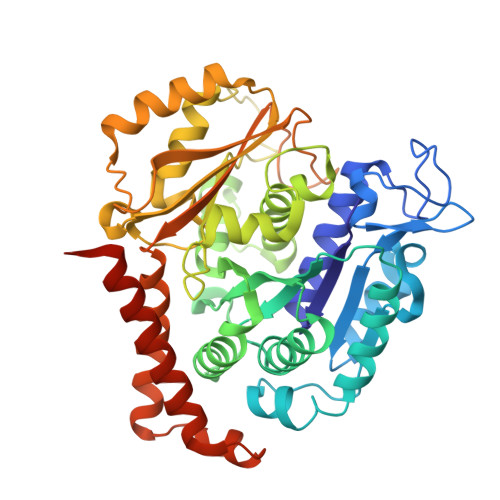
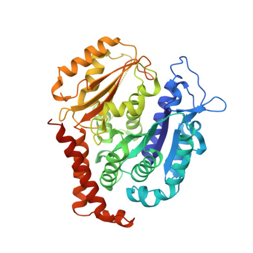
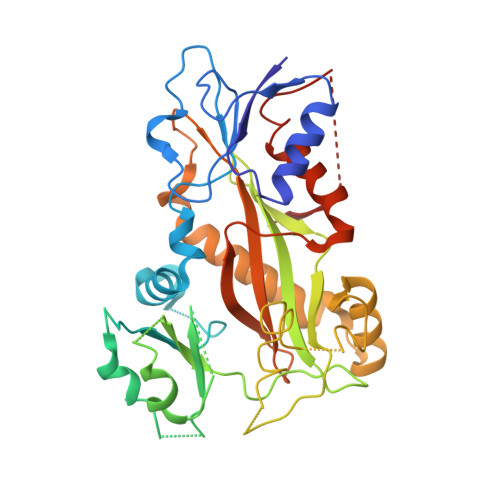
Design, synthesis, biological evaluation and cocrystal structures with tubulin of chiral beta-lactam bridged combretastatin A-4 analogues as potent antitumor agents

Zhou, P.Liang, Y.Zhang, H.Jiang, H.Feng, K.Xu, P.Wang, J.Wang, X.Ding, K.Luo, C.Liu, M.Wang, Y.

(2017) Eur J Med Chem 144: 817-842

  • DOI: https://doi.org/10.1016/j.ejmech.2017.12.004
  • Primary Citation Related Structures: 
    5XAF, 5XAG

  • PubMed Abstract: 

    A diverse of chiral β-lactam bridged analogues of combretastatin A-4 (CA-4), 3-substituted 1,4-diaryl-2-azetidinones, were asymmetrically synthesized and biologically evaluated, leading to identify a number of potent anti-proliferative compounds represented by 14b and 14c with IC 50 values of 0.001-0.021 μM, against four human cancer cell lines (A2780, Hela, SKOV-3 and MDA-MB-231). Structure-activity relationship (SAR) studies on all stereoisomers of 14b and 14c revealed that the absolute configurations of the chiral centers at 3- and 4-position were critically important for the activity and generally a trans configuration between the "A" and "B" rings is optimal. In addition, 14b and 14c displayed less cytotoxicity on normal human oviduct epithelial cells than malignant cells indicating good selectivity in vitro. Further biochemical evaluation and cocrystal structures with tubulin demonstrated that both compounds disrupted tubulin polymerization through interacting at the colchicine-binding site, suppressed angiogenesis in vitro and in vivo, blocked cell cycle progression at mitotic phase and induced cellular apoptosis. The in vivo assays verified that both compounds inhibited xenograft tumor growth in nude mice with acceptable therapeutic window, showing promising potentials for further clinical development.


  • Organizational Affiliation
    • School of Pharmacy, Fudan University, Shanghai 201203, China; State Key Laboratory of Organometallic Chemistry, Shanghai Institute of Organic Chemistry, Chinese Academy of Sciences, Shanghai 200032, China.

Macromolecule Content 

  • Total Structure Weight: 271.02 kDa 
  • Atom Count: 18,303 
  • Modeled Residue Count: 2,159 
  • Deposited Residue Count: 2,359 
  • Unique protein chains: 4

Macromolecules

Find similar proteins by:|  3D Structure
Entity ID: 1
MoleculeChains  Sequence LengthOrganismDetailsImage
Tubulin alpha-1B chain
A, C
451Bos taurusMutation(s): 0 
EC: 3.6.5
UniProt
Find proteins for P81947 (Bos taurus)
Explore P81947 
Go to UniProtKB:  P81947
Entity Groups
Sequence Clusters30% Identity50% Identity70% Identity90% Identity95% Identity100% Identity
UniProt GroupP81947
Sequence Annotations
Expand
Reference Sequence
Find similar proteins by:|  3D Structure
Entity ID: 2
MoleculeChains  Sequence LengthOrganismDetailsImage
Tubulin beta-2B chain
B, D
445Bos taurusMutation(s): 0 
Gene Names: TUBB2B
UniProt
Find proteins for Q6B856 (Bos taurus)
Explore Q6B856 
Go to UniProtKB:  Q6B856
Entity Groups
Sequence Clusters30% Identity50% Identity70% Identity90% Identity95% Identity100% Identity
UniProt GroupQ6B856
Sequence Annotations
Expand
Reference Sequence
Find similar proteins by:|  3D Structure
Entity ID: 3
MoleculeChains  Sequence LengthOrganismDetailsImage
Stathmin-4189Rattus norvegicusMutation(s): 0 
Gene Names: Stmn4
UniProt
Find proteins for P63043 (Rattus norvegicus)
Explore P63043 
Go to UniProtKB:  P63043
Entity Groups
Sequence Clusters30% Identity50% Identity70% Identity90% Identity95% Identity100% Identity
UniProt GroupP63043
Sequence Annotations
Expand
Reference Sequence
Find similar proteins by:|  3D Structure
Entity ID: 4
MoleculeChains  Sequence LengthOrganismDetailsImage
TTL Protein378Gallus gallusMutation(s): 0 
Gene Names: TTL

Small Molecules

Ligands 9 Unique
IDChains Name / Formula / InChI Key2D Diagram3D Interactions
GTP

Query on GTP


Download:Ideal Coordinates CCD File
G [auth A],
V [auth C]
GUANOSINE-5'-TRIPHOSPHATE
C10 H16 N5 O14 P3
XKMLYUALXHKNFT-UUOKFMHZSA-N
ACP

Query on ACP


Download:Ideal Coordinates CCD File
KA [auth F]PHOSPHOMETHYLPHOSPHONIC ACID ADENYLATE ESTER
C11 H18 N5 O12 P3
UFZTZBNSLXELAL-IOSLPCCCSA-N
GDP

Query on GDP


Download:Ideal Coordinates CCD File
DA [auth D],
M [auth B]
GUANOSINE-5'-DIPHOSPHATE
C10 H15 N5 O11 P2
QGWNDRXFNXRZMB-UUOKFMHZSA-N
84F

Query on 84F


Download:Ideal Coordinates CCD File
HA [auth D],
T [auth B]
(3S,4R)-4-(3-hydroxy-4-methoxyphenyl)-3-methyl-1-(3,4,5-trimethoxyphenyl)azetidin-2-one
C20 H23 N O6
KLFAJLHIJQPMNJ-BBATYDOGSA-N
MES

Query on MES


Download:Ideal Coordinates CCD File
S [auth B]2-(N-MORPHOLINO)-ETHANESULFONIC ACID
C6 H13 N O4 S
SXGZJKUKBWWHRA-UHFFFAOYSA-N
GOL

Query on GOL


Download:Ideal Coordinates CCD File
FA [auth D]
GA [auth D]
I [auth A]
J [auth A]
L [auth A]
FA [auth D],
GA [auth D],
I [auth A],
J [auth A],
L [auth A],
O [auth B],
P [auth B],
Q [auth B],
U [auth C],
X [auth C],
Y [auth C],
Z [auth C]
GLYCEROL
C3 H8 O3
PEDCQBHIVMGVHV-UHFFFAOYSA-N
IMD

Query on IMD


Download:Ideal Coordinates CCD File
BA [auth C],
CA [auth C],
IA [auth E]
IMIDAZOLE
C3 H5 N2
RAXXELZNTBOGNW-UHFFFAOYSA-O
CA

Query on CA


Download:Ideal Coordinates CCD File
AA [auth C],
K [auth A],
R [auth B]
CALCIUM ION
Ca
BHPQYMZQTOCNFJ-UHFFFAOYSA-N
MG

Query on MG


Download:Ideal Coordinates CCD File
EA [auth D],
H [auth A],
JA [auth F],
N [auth B],
W [auth C]
MAGNESIUM ION
Mg
JLVVSXFLKOJNIY-UHFFFAOYSA-N

Experimental Data & Validation

Experimental Data

  • Method: X-RAY DIFFRACTION
  • Resolution: 2.55 Å
  • R-Value Free:  0.257 (Depositor), 0.258 (DCC) 
  • R-Value Work:  0.206 (Depositor), 0.210 (DCC) 
  • R-Value Observed: 0.207 (Depositor) 
Space Group: P 21 21 21
Unit Cell:
Length ( Å )Angle ( ˚ )
a = 104.568α = 90
b = 156.509β = 90
c = 182.078γ = 90
Software Package:
Software NamePurpose
PHENIXrefinement
XDSdata reduction
Aimlessdata scaling
PHASERphasing

Structure Validation

View Full Validation Report


Ligand Structure Quality Assessment 


Entry History 

& Funding Information

Deposition Data


Funding OrganizationLocationGrant Number
China--

Revision History  (Full details and data files)

  • Version 1.0: 2017-12-20
    Type: Initial release
  • Version 1.1: 2018-01-24
    Changes: Database references
  • Version 1.2: 2023-11-22
    Changes: Data collection, Database references, Derived calculations, Refinement description