5XA1 | pdb_00005xa1

Crystal structure of inositol 1,4,5-trisphosphate receptor cytosolic domain with inositol 1,4,5-trisphosphate


Experimental Data Snapshot

  • Method: X-RAY DIFFRACTION
  • Resolution: 6.20 Å
  • R-Value Free: 
    0.382 (Depositor), 0.390 (DCC) 
  • R-Value Work: 
    0.360 (Depositor), 0.352 (DCC) 
  • R-Value Observed: 
    0.361 (Depositor) 

Starting Models: experimental
View more details

wwPDB Validation 3D Report Full Report

Validation slider image for 5XA1

Ligand Structure Quality Assessment 


This is version 1.2 of the entry. See complete history

Literature

IP3-mediated gating mechanism of the IP3 receptor revealed by mutagenesis and X-ray crystallography

Hamada, K.Miyatake, H.Terauchi, A.Mikoshiba, K.

(2017) Proc Natl Acad Sci U S A 114: 4661-4666

  • DOI: https://doi.org/10.1073/pnas.1701420114
  • Primary Citation Related Structures: 
    5GUG, 5X9Z, 5XA0, 5XA1

  • PubMed Abstract: 

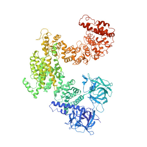
    The inositol 1,4,5-trisphosphate (IP 3 ) receptor (IP 3 R) is an IP 3 -gated ion channel that releases calcium ions (Ca 2+ ) from the endoplasmic reticulum. The IP 3 -binding sites in the large cytosolic domain are distant from the Ca 2+ conducting pore, and the allosteric mechanism of how IP 3 opens the Ca 2+ channel remains elusive. Here, we identify a long-range gating mechanism uncovered by channel mutagenesis and X-ray crystallography of the large cytosolic domain of mouse type 1 IP 3 R in the absence and presence of IP 3 Analyses of two distinct space group crystals uncovered an IP 3 -dependent global translocation of the curvature α-helical domain interfacing with the cytosolic and channel domains. Mutagenesis of the IP 3 R channel revealed an essential role of a leaflet structure in the α-helical domain. These results suggest that the curvature α-helical domain relays IP 3 -controlled global conformational dynamics to the channel through the leaflet, conferring long-range allosteric coupling from IP 3 binding to the Ca 2+ channel.


  • Organizational Affiliation
    • Laboratory for Developmental Neurobiology, Brain Science Institute, RIKEN, Saitama 351-0198, Japan.

Macromolecule Content 

  • Total Structure Weight: 362.86 kDa 
  • Atom Count: 15,198 
  • Modeled Residue Count: 2,514 
  • Deposited Residue Count: 3,162 
  • Unique protein chains: 1

Macromolecules

Find similar proteins by:|  3D Structure
Entity ID: 1
MoleculeChains  Sequence LengthOrganismDetailsImage
Inositol 1,4,5-trisphosphate receptor type 1
A, B
1,581Mus musculusMutation(s): 0 
Gene Names: Itpr1Insp3rPcd6Pcp1
UniProt & NIH Common Fund Data Resources
Find proteins for P11881 (Mus musculus)
Explore P11881 
Go to UniProtKB:  P11881
IMPC:  MGI:96623
Entity Groups
Sequence Clusters30% Identity50% Identity70% Identity90% Identity95% Identity100% Identity
UniProt GroupP11881
Sequence Annotations
Expand
Reference Sequence

Small Molecules

Binding Affinity Annotations 
IDSourceBinding Affinity
I3P BindingDB:  5XA1 Kd: min: 0.21, max: 260 (nM) from 5 assay(s)
IC50: 300 (nM) from 1 assay(s)
EC50: min: 20, max: 209 (nM) from 7 assay(s)

Experimental Data & Validation

Experimental Data

  • Method: X-RAY DIFFRACTION
  • Resolution: 6.20 Å
  • R-Value Free:  0.382 (Depositor), 0.390 (DCC) 
  • R-Value Work:  0.360 (Depositor), 0.352 (DCC) 
  • R-Value Observed: 0.361 (Depositor) 
Space Group: P 42
Unit Cell:
Length ( Å )Angle ( ˚ )
a = 126.803α = 90
b = 126.803β = 90
c = 367.784γ = 90
Software Package:
Software NamePurpose
PHENIXrefinement
XDSdata reduction
XDSdata scaling
PHENIXphasing

Structure Validation

View Full Validation Report


Ligand Structure Quality Assessment 


Entry History 

& Funding Information

Deposition Data


Funding OrganizationLocationGrant Number
JSPSJapan15K06791

Revision History  (Full details and data files)

  • Version 1.0: 2017-04-19
    Type: Initial release
  • Version 1.1: 2017-05-17
    Changes: Database references
  • Version 1.2: 2023-11-22
    Changes: Data collection, Database references, Refinement description