5X9W

Mismatch Repair Protein


Experimental Data Snapshot

  • Method: X-RAY DIFFRACTION
  • Resolution: 3.30 Å
  • R-Value Free: 0.273 
  • R-Value Work: 0.221 
  • R-Value Observed: 0.224 

wwPDB Validation   3D Report Full Report


Ligand Structure Quality Assessment 


This is version 1.3 of the entry. See complete history


Literature

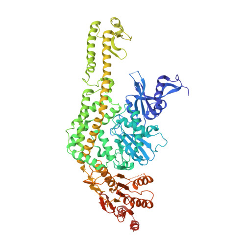
Mechanism of formation of a toroid around DNA by the mismatch sensor protein.

Nirwal, S.Kulkarni, D.S.Sharma, A.Rao, D.N.Nair, D.T.

(2018) Nucleic Acids Res 46: 256-266

  • DOI: https://doi.org/10.1093/nar/gkx1149
  • Primary Citation of Related Structures:  
    5X9W, 5YK4

  • PubMed Abstract: 

    The DNA mismatch repair (MMR) pathway removes errors that appear during genome replication. MutS is the primary mismatch sensor and forms an asymmetric dimer that encircles DNA to bend it to scan for mismatches. The mechanism utilized to load DNA into the central tunnel was unknown and the origin of the force required to bend DNA was unclear. We show that, in absence of DNA, MutS forms a symmetric dimer wherein a gap exists between the monomers through which DNA can enter the central tunnel. The comparison with structures of MutS-DNA complexes suggests that the mismatch scanning monomer (Bm) will move by nearly 50 Å to associate with the other monomer (Am). Consequently, the N-terminal domains of both monomers will press onto DNA to bend it. The proposed mechanism of toroid formation evinces that the force required to bend DNA arises primarily due to the movement of Bm and hence, the MutS dimer acts like a pair of pliers to bend DNA. We also shed light on the allosteric mechanism that influences the expulsion of adenosine triphosphate from Am on DNA binding. Overall, this study provides mechanistic insight regarding the primary event in MMR i.e. the assembly of the MutS-DNA complex.


  • Organizational Affiliation

    Regional Centre for Biotechnology, NCR Biotech Science Cluster, 3rd Milestone, Faridabad-Gurgaon Expressway, Faridabad, 121001, Haryana, India.


Macromolecules
Find similar proteins by:  (by identity cutoff)  |  3D Structure
Entity ID: 1
MoleculeChains Sequence LengthOrganismDetailsImage
DNA mismatch repair protein MutS
A, B
816Neisseria gonorrhoeae FA 1090Mutation(s): 0 
Gene Names: mutSNGO1930
UniProt
Find proteins for Q5F5J4 (Neisseria gonorrhoeae (strain ATCC 700825 / FA 1090))
Explore Q5F5J4 
Go to UniProtKB:  Q5F5J4
Entity Groups  
Sequence Clusters30% Identity50% Identity70% Identity90% Identity95% Identity100% Identity
UniProt GroupQ5F5J4
Sequence Annotations
Expand
  • Reference Sequence
Small Molecules
Ligands 1 Unique
IDChains Name / Formula / InChI Key2D Diagram3D Interactions
ANP
Query on ANP

Download Ideal Coordinates CCD File 
C [auth A],
D [auth B]
PHOSPHOAMINOPHOSPHONIC ACID-ADENYLATE ESTER
C10 H17 N6 O12 P3
PVKSNHVPLWYQGJ-KQYNXXCUSA-N
Experimental Data & Validation

Experimental Data

  • Method: X-RAY DIFFRACTION
  • Resolution: 3.30 Å
  • R-Value Free: 0.273 
  • R-Value Work: 0.221 
  • R-Value Observed: 0.224 
  • Space Group: P 21 2 21
Unit Cell:
Length ( Å )Angle ( ˚ )
a = 89.51α = 90
b = 102.07β = 90
c = 235.72γ = 90
Software Package:
Software NamePurpose
REFMACrefinement
iMOSFLMdata reduction
SCALAdata scaling
PHASERphasing

Structure Validation

View Full Validation Report


Ligand Structure Quality Assessment 


Entry History & Funding Information

Deposition Data


Funding OrganizationLocationGrant Number
Department of BiotechnologyIndia--

Revision History  (Full details and data files)

  • Version 1.0: 2017-11-22
    Type: Initial release
  • Version 1.1: 2017-12-13
    Changes: Database references
  • Version 1.2: 2018-01-24
    Changes: Database references
  • Version 1.3: 2023-11-22
    Changes: Data collection, Database references, Refinement description