5WZY

Crystal structure of the P2X4 receptor from zebrafish in the presence of CTP at 2.8 Angstroms


Experimental Data Snapshot

  • Method: X-RAY DIFFRACTION
  • Resolution: 2.80 Å
  • R-Value Free: 0.249 
  • R-Value Work: 0.213 
  • R-Value Observed: 0.216 

wwPDB Validation   3D Report Full Report


Ligand Structure Quality Assessment 


This is version 2.0 of the entry. See complete history


Literature

Structural insights into the nucleotide base specificity of P2X receptors

Kasuya, G.Fujiwara, Y.Tsukamoto, H.Morinaga, S.Ryu, S.Touhara, K.Ishitani, R.Furutani, Y.Hattori, M.Nureki, O.

(2017) Sci Rep 7: 45208-45208

  • DOI: https://doi.org/10.1038/srep45208
  • Primary Citation of Related Structures:  
    5WZY

  • PubMed Abstract: 

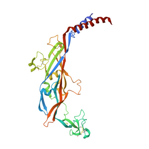
    P2X receptors are trimeric ATP-gated cation channels involved in diverse physiological processes, ranging from muscle contraction to nociception. Despite the recent structure determination of the ATP-bound P2X receptors, the molecular mechanism of the nucleotide base specificity has remained elusive. Here, we present the crystal structure of zebrafish P2X4 in complex with a weak affinity agonist, CTP, together with structure-based electrophysiological and spectroscopic analyses. The CTP-bound structure revealed a hydrogen bond, between the cytosine base and the side chain of the basic residue in the agonist binding site, which mediates the weak but significant affinity for CTP. The cytosine base is further recognized by two main chain atoms, as in the ATP-bound structure, but their bond lengths seem to be extended in the CTP-bound structure, also possibly contributing to the weaker affinity for CTP over ATP. This work provides the structural insights for the nucleotide base specificity of P2X receptors.


  • Organizational Affiliation

    Department of Biological Sciences, Graduate School of Science, The University of Tokyo, 2-11-16 Yayoi, Bunkyo-ku, Tokyo, 113-0032, Japan.


Macromolecules
Find similar proteins by:  (by identity cutoff)  |  3D Structure
Entity ID: 1
MoleculeChains Sequence LengthOrganismDetailsImage
p2X purinoceptor340Danio rerioMutation(s): 0 
Gene Names: p2rx4a
Membrane Entity: Yes 
UniProt
Find proteins for F8W463 (Danio rerio)
Explore F8W463 
Go to UniProtKB:  F8W463
Entity Groups  
Sequence Clusters30% Identity50% Identity70% Identity90% Identity95% Identity100% Identity
UniProt GroupF8W463
Sequence Annotations
Expand
  • Reference Sequence
Oligosaccharides

Help

Entity ID: 2
MoleculeChains Length2D Diagram Glycosylation3D Interactions
alpha-D-mannopyranose-(1-3)-alpha-D-mannopyranose-(1-3)-[alpha-D-mannopyranose-(1-6)]beta-D-mannopyranose-(1-4)-2-acetamido-2-deoxy-beta-D-glucopyranose-(1-4)-2-acetamido-2-deoxy-beta-D-glucopyranose
B
6N-Glycosylation
Glycosylation Resources
GlyTouCan:  G42984ZU
GlyCosmos:  G42984ZU
GlyGen:  G42984ZU
Experimental Data & Validation

Experimental Data

  • Method: X-RAY DIFFRACTION
  • Resolution: 2.80 Å
  • R-Value Free: 0.249 
  • R-Value Work: 0.213 
  • R-Value Observed: 0.216 
  • Space Group: H 3 2
Unit Cell:
Length ( Å )Angle ( ˚ )
a = 129.242α = 90
b = 129.242β = 90
c = 251.825γ = 120
Software Package:
Software NamePurpose
HKL-2000data scaling
PHENIXrefinement
PDB_EXTRACTdata extraction
HKL-2000data reduction
PHASERphasing
HKLdata scaling

Structure Validation

View Full Validation Report


Ligand Structure Quality Assessment 


Entry History & Funding Information

Deposition Data


Funding OrganizationLocationGrant Number
Japan Society for the Promotion of ScienceJapan24227004
National Natural Science Foundation of ChinaChina31650110469
National Natural Science Foundation of ChinaChina31570838
Ministry of Science and Technology of ChinaChina2016YFA0502800

Revision History  (Full details and data files)

  • Version 1.0: 2017-04-05
    Type: Initial release
  • Version 1.1: 2020-02-26
    Changes: Data collection
  • Version 2.0: 2020-07-29
    Type: Remediation
    Reason: Carbohydrate remediation
    Changes: Atomic model, Data collection, Derived calculations, Structure summary