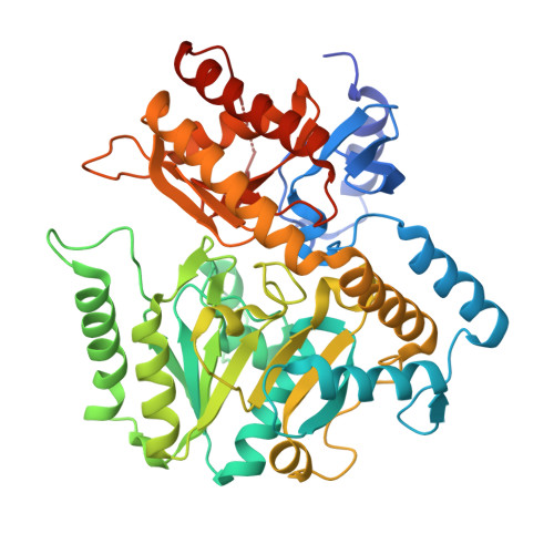
5WYF

Structure of amino acid racemase, 2.12 A


Experimental Data Snapshot

  • Method: X-RAY DIFFRACTION
  • Resolution: 2.12 Å
  • R-Value Free: 0.221 
  • R-Value Work: 0.176 
  • R-Value Observed: 0.178 

wwPDB Validation   3D Report Full Report


Ligand Structure Quality Assessment 


This is version 1.2 of the entry. See complete history


Literature

Crystal structure of the novel amino-acid racemase isoleucine 2-epimerase from Lactobacillus buchneri.

Hayashi, J.Mutaguchi, Y.Minemura, Y.Nakagawa, N.Yoneda, K.Ohmori, T.Ohshima, T.Sakuraba, H.

(2017) Acta Crystallogr D Struct Biol 73: 428-437

  • DOI: https://doi.org/10.1107/S2059798317005332
  • Primary Citation of Related Structures:  
    4YSN, 4YSV, 5WYA, 5WYF

  • PubMed Abstract: 

    Crystal structures of Lactobacillus buchneri isoleucine 2-epimerase, a novel branched-chain amino-acid racemase, were determined for the enzyme in the apo form, in complex with pyridoxal 5'-phosphate (PLP), in complex with N-(5'-phosphopyridoxyl)-L-isoleucine (PLP-L-Ile) and in complex with N-(5'-phosphopyridoxyl)-D-allo-isoleucine (PLP-D-allo-Ile) at resolutions of 2.77, 1.94, 2.65 and 2.12 Å, respectively. The enzyme assembled as a tetramer, with each subunit being composed of N-terminal, C-terminal and large PLP-binding domains. The active-site cavity in the apo structure was much more solvent-accessible than that in the PLP-bound structure. This indicates that a marked structural change occurs around the active site upon binding of PLP that provides a solvent-inaccessible environment for the enzymatic reaction. The main-chain coordinates of the L. buchneri isoleucine 2-epimerase monomer showed a notable similarity to those of α-amino-ℇ-caprolactam racemase from Achromobactor obae and γ-aminobutyrate aminotransferase from Escherichia coli. However, the amino-acid residues involved in substrate binding in those two enzymes are only partially conserved in L. buchneri isoleucine 2-epimerase, which may account for the differences in substrate recognition by the three enzymes. The structures bound with reaction-intermediate analogues (PLP-L-Ile and PLP-D-allo-Ile) and site-directed mutagenesis suggest that L-isoleucine epimerization proceeds through abstraction of the α-hydrogen of the substrate by Lys280, while Asp222 serves as the catalytic residue adding an α-hydrogen to the quinonoid intermediate to form D-allo-isoleucine.


  • Organizational Affiliation

    Department of Applied Biological Science, Faculty of Agriculture, Kagawa University, 2393 Ikenobe, Miki-cho, Kita-gun, Kagawa 761-0795, Japan.


Macromolecules
Find similar proteins by:  (by identity cutoff)  |  3D Structure
Entity ID: 1
MoleculeChains Sequence LengthOrganismDetailsImage
Isoleucine 2-epimerase
A, B, C, D
462Lentilactobacillus buchneriMutation(s): 0 
EC: 5.1.1.21
UniProt
Find proteins for M1GRN3 (Lentilactobacillus buchneri)
Explore M1GRN3 
Go to UniProtKB:  M1GRN3
Entity Groups  
Sequence Clusters30% Identity50% Identity70% Identity90% Identity95% Identity100% Identity
UniProt GroupM1GRN3
Sequence Annotations
Expand
  • Reference Sequence
Small Molecules
Ligands 2 Unique
IDChains Name / Formula / InChI Key2D Diagram3D Interactions
ILP
Query on ILP

Download Ideal Coordinates CCD File 
E [auth A],
JA [auth D],
P [auth B],
Y [auth C]
N-[O-PHOSPHONO-PYRIDOXYL]-ISOLEUCINE
C14 H23 N2 O7 P
GZZDWFDWHXPWJK-QPUJVOFHSA-N
CD
Query on CD

Download Ideal Coordinates CCD File 
AA [auth C]
BA [auth C]
CA [auth C]
DA [auth C]
EA [auth C]
AA [auth C],
BA [auth C],
CA [auth C],
DA [auth C],
EA [auth C],
F [auth A],
FA [auth C],
G [auth A],
GA [auth C],
H [auth A],
HA [auth C],
I [auth A],
IA [auth C],
J [auth A],
K [auth A],
KA [auth D],
L [auth A],
LA [auth D],
M [auth A],
MA [auth D],
N [auth A],
NA [auth D],
O [auth A],
OA [auth D],
PA [auth D],
Q [auth B],
QA [auth D],
R [auth B],
RA [auth D],
S [auth B],
T [auth B],
U [auth B],
V [auth B],
W [auth B],
X [auth B],
Z [auth C]
CADMIUM ION
Cd
WLZRMCYVCSSEQC-UHFFFAOYSA-N
Experimental Data & Validation

Experimental Data

  • Method: X-RAY DIFFRACTION
  • Resolution: 2.12 Å
  • R-Value Free: 0.221 
  • R-Value Work: 0.176 
  • R-Value Observed: 0.178 
  • Space Group: P 1 21 1
Unit Cell:
Length ( Å )Angle ( ˚ )
a = 59.514α = 90
b = 85.277β = 96.93
c = 186.566γ = 90
Software Package:
Software NamePurpose
HKL-2000data scaling
MOLREPphasing
REFMACrefinement
PDB_EXTRACTdata extraction
HKL-2000data reduction

Structure Validation

View Full Validation Report


Ligand Structure Quality Assessment 


Entry History 

Deposition Data

Revision History  (Full details and data files)

  • Version 1.0: 2017-04-19
    Type: Initial release
  • Version 1.1: 2017-05-17
    Changes: Database references
  • Version 1.2: 2023-11-22
    Changes: Data collection, Database references, Derived calculations, Refinement description