5WRO | pdb_00005wro

Crystal structure of Drosophila enolase


Experimental Data Snapshot

  • Method: X-RAY DIFFRACTION
  • Resolution: 2.02 Å
  • R-Value Free: 
    0.200 (Depositor), 0.198 (DCC) 
  • R-Value Work: 
    0.159 (Depositor), 0.160 (DCC) 
  • R-Value Observed: 
    0.161 (Depositor) 

Starting Model: experimental
View more details

wwPDB Validation 3D Report Full Report

Validation slider image for 5WRO

This is version 1.2 of the entry. See complete history

Literature

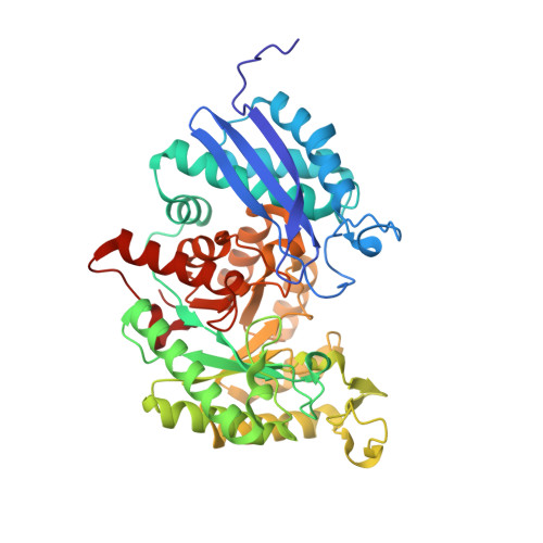
Crystal structure of enolase from Drosophila melanogaster.

Sun, C.Xu, B.Liu, X.Zhang, Z.Su, Z.

(2017) Acta Crystallogr F Struct Biol Commun 73: 228-234

  • DOI: https://doi.org/10.1107/S2053230X17004022
  • Primary Citation Related Structures: 
    5WRO

  • PubMed Abstract: 

    Enolase is an important enzyme in glycolysis and various biological processes. Its dysfunction is closely associated with diseases. Here, the enolase from Drosophila melanogaster (DmENO) was purified and crystallized. A crystal of DmENO diffracted to 2.0 Å resolution and belonged to space group R32. The structure was solved by molecular replacement. Like most enolases, DmENO forms a homodimer with conserved residues in the dimer interface. DmENO possesses an open conformation in this structure and contains conserved elements for catalytic activity. This work provides a structural basis for further functional and evolutionary studies of enolase.


  • Organizational Affiliation
    • College of Chemical Engineering, Qingdao University of Science and Technology, Qingdao, Shandong 266042, People's Republic of China.

Macromolecule Content 

  • Total Structure Weight: 48.03 kDa 
  • Atom Count: 3,664 
  • Modeled Residue Count: 439 
  • Deposited Residue Count: 439 
  • Unique protein chains: 1

Macromolecules

Find similar proteins by:|  3D Structure
Entity ID: 1
MoleculeChains  Sequence LengthOrganismDetailsImage
Enolase439Drosophila melanogasterMutation(s): 0 
Gene Names: EnoCG17654
EC: 4.2.1.11
UniProt
Find proteins for P15007 (Drosophila melanogaster)
Explore P15007 
Go to UniProtKB:  P15007
Entity Groups
Sequence Clusters30% Identity50% Identity70% Identity90% Identity95% Identity100% Identity
UniProt GroupP15007
Sequence Annotations
Expand
Reference Sequence

Small Molecules

Ligands 5 Unique
IDChains Name / Formula / InChI Key2D Diagram3D Interactions
CD

Query on CD


Download:Ideal Coordinates CCD File
B [auth A],
C [auth A]
CADMIUM ION
Cd
WLZRMCYVCSSEQC-UHFFFAOYSA-N
SO4

Query on SO4


Download:Ideal Coordinates CCD File
F [auth A],
G [auth A]
SULFATE ION
O4 S
QAOWNCQODCNURD-UHFFFAOYSA-L
GOL

Query on GOL


Download:Ideal Coordinates CCD File
H [auth A],
I [auth A]
GLYCEROL
C3 H8 O3
PEDCQBHIVMGVHV-UHFFFAOYSA-N
CO

Query on CO


Download:Ideal Coordinates CCD File
D [auth A]COBALT (II) ION
Co
XLJKHNWPARRRJB-UHFFFAOYSA-N
CL

Query on CL


Download:Ideal Coordinates CCD File
E [auth A]CHLORIDE ION
Cl
VEXZGXHMUGYJMC-UHFFFAOYSA-M

Experimental Data & Validation

Experimental Data

  • Method: X-RAY DIFFRACTION
  • Resolution: 2.02 Å
  • R-Value Free:  0.200 (Depositor), 0.198 (DCC) 
  • R-Value Work:  0.159 (Depositor), 0.160 (DCC) 
  • R-Value Observed: 0.161 (Depositor) 
Space Group: H 3 2
Unit Cell:
Length ( Å )Angle ( ˚ )
a = 118.82α = 90
b = 118.82β = 90
c = 229.792γ = 120
Software Package:
Software NamePurpose
PHENIXrefinement
HKL-2000data reduction
HKL-2000data scaling
PHASERphasing

Structure Validation

View Full Validation Report


Entry History 

Deposition Data

Revision History  (Full details and data files)

  • Version 1.0: 2017-04-26
    Type: Initial release
  • Version 1.1: 2023-11-08
    Changes: Data collection, Database references, Derived calculations, Refinement description
  • Version 1.2: 2024-10-09
    Changes: Structure summary