5WQ7

CryoEM structure of type II secretion system secretin GspD in E.coli K12


Experimental Data Snapshot

  • Method: ELECTRON MICROSCOPY
  • Resolution: 3.04 Å
  • Aggregation State: PARTICLE 
  • Reconstruction Method: SINGLE PARTICLE 

wwPDB Validation   3D Report Full Report


This is version 1.4 of the entry. See complete history


Literature

Structural insights into the secretin translocation channel in the type II secretion system

Yan, Z.Yin, M.Xu, D.Zhu, Y.Li, X.

(2017) Nat Struct Mol Biol 24: 177-183

  • DOI: https://doi.org/10.1038/nsmb.3350
  • Primary Citation of Related Structures:  
    5WQ7, 5WQ8, 5WQ9

  • PubMed Abstract: 

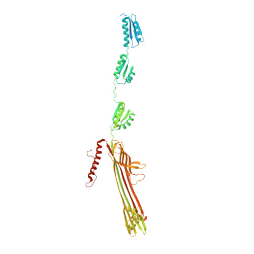
    The secretin GspD of the type II secretion system (T2SS) forms a channel across the outer membrane in Gram-negative bacteria to transport substrates from the periplasm to the extracellular milieu. The lack of an atomic-resolution structure of the GspD channel hinders the investigation of substrate translocation mechanism of T2SS. Here we report cryo-EM structures of two GspD channels (∼1 MDa), from Escherichia coli K12 and Vibrio cholerae, at ∼3 Å resolution. The structures reveal a pentadecameric channel architecture, wherein three rings of GspD N domains form the periplasmic channel. The secretin domain constitutes a novel double β-barrel channel, with at least 60 β-strands in each barrel, and is stabilized by S domains. The outer membrane channel is sealed by β-strand-enriched gates. On the basis of the partially open state captured, we proposed a detailed gate-opening mechanism. Our structures provide a structural basis for understanding the secretin superfamily and the mechanism of substrate translocation in T2SS.


  • Organizational Affiliation

    School of Life Sciences, Tsinghua University, Beijing, China.


Macromolecules
Find similar proteins by:  (by identity cutoff)  |  3D Structure
Entity ID: 1
MoleculeChains Sequence LengthOrganismDetailsImage
Putative type II secretion system protein D
A, B, C, D, E
A, B, C, D, E, F, G, H, I, J, K, L, M, N, O
627Escherichia coli K-12Mutation(s): 0 
Gene Names: gspDyheFb3325JW5707
Membrane Entity: Yes 
UniProt
Find proteins for P45758 (Escherichia coli (strain K12))
Explore P45758 
Go to UniProtKB:  P45758
Entity Groups  
Sequence Clusters30% Identity50% Identity70% Identity90% Identity95% Identity100% Identity
UniProt GroupP45758
Sequence Annotations
Expand
  • Reference Sequence
Experimental Data & Validation

Experimental Data

  • Method: ELECTRON MICROSCOPY
  • Resolution: 3.04 Å
  • Aggregation State: PARTICLE 
  • Reconstruction Method: SINGLE PARTICLE 
EM Software:
TaskSoftware PackageVersion
RECONSTRUCTIONRELION

Structure Validation

View Full Validation Report


Entry History 

Deposition Data

Revision History  (Full details and data files)

  • Version 1.0: 2016-12-28
    Type: Initial release
  • Version 1.1: 2017-01-11
    Changes: Database references
  • Version 1.2: 2017-02-22
    Changes: Database references
  • Version 1.3: 2019-10-16
    Changes: Data collection, Other
  • Version 1.4: 2024-03-27
    Changes: Data collection, Database references