5WO9

Crystal Structure of Transient Receptor Potential (TRP) channel TRPV6* in the presence of Calcium


Experimental Data Snapshot

  • Method: X-RAY DIFFRACTION
  • Resolution: 3.70 Å
  • R-Value Free: 0.316 
  • R-Value Work: 0.272 
  • R-Value Observed: 0.274 

wwPDB Validation   3D Report Full Report


Ligand Structure Quality Assessment 


This is version 1.3 of the entry. See complete history


Literature

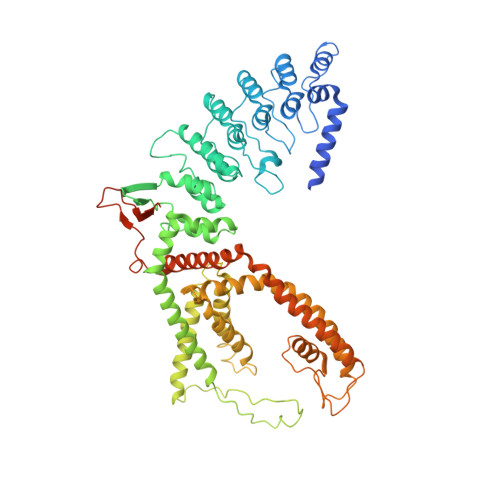
Swapping of transmembrane domains in the epithelial calcium channel TRPV6.

Singh, A.K.Saotome, K.Sobolevsky, A.I.

(2017) Sci Rep 7: 10669-10669

  • DOI: https://doi.org/10.1038/s41598-017-10993-9
  • Primary Citation of Related Structures:  
    5WO6, 5WO7, 5WO8, 5WO9, 5WOA

  • PubMed Abstract: 

    Tetrameric ion channels have either swapped or non-swapped arrangements of the S1-S4 and pore domains. Here we show that mutations in the transmembrane domain of TRPV6 can result in conversion from a domain-swapped to non-swapped fold. These results reveal structural determinants of domain swapping and raise the possibility that a single ion channel subtype can fold into either arrangement in vivo, affecting its function in normal or disease states.


  • Organizational Affiliation

    Department of Biochemistry and Molecular Biophysics, Columbia University 650 West 168th Street, New York, NY, 10032, USA.


Macromolecules
Find similar proteins by:  (by identity cutoff)  |  3D Structure
Entity ID: 1
MoleculeChains Sequence LengthOrganismDetailsImage
Transient receptor potential cation channel subfamily V member 6672Rattus norvegicusMutation(s): 3 
Gene Names: Trpv6
Membrane Entity: Yes 
UniProt
Find proteins for Q9R186 (Rattus norvegicus)
Explore Q9R186 
Go to UniProtKB:  Q9R186
Entity Groups  
Sequence Clusters30% Identity50% Identity70% Identity90% Identity95% Identity100% Identity
UniProt GroupQ9R186
Sequence Annotations
Expand
  • Reference Sequence
Experimental Data & Validation

Experimental Data

  • Method: X-RAY DIFFRACTION
  • Resolution: 3.70 Å
  • R-Value Free: 0.316 
  • R-Value Work: 0.272 
  • R-Value Observed: 0.274 
  • Space Group: P 4 21 2
Unit Cell:
Length ( Å )Angle ( ˚ )
a = 145.48α = 90
b = 145.48β = 90
c = 110.95γ = 90
Software Package:
Software NamePurpose
PHENIXrefinement
XDSdata reduction
XDSdata scaling
PHASERphasing

Structure Validation

View Full Validation Report


Ligand Structure Quality Assessment 


Entry History & Funding Information

Deposition Data


Funding OrganizationLocationGrant Number
National Institutes of Health/National Cancer Institute (NIH/NCI)United StatesR01 CA206573

Revision History  (Full details and data files)

  • Version 1.0: 2017-09-13
    Type: Initial release
  • Version 1.1: 2017-09-27
    Changes: Author supporting evidence, Database references
  • Version 1.2: 2019-12-04
    Changes: Author supporting evidence
  • Version 1.3: 2023-10-04
    Changes: Data collection, Database references, Refinement description