5WMU

Structural Insights into Substrate and Inhibitor Binding Sites in Human Indoleamine 2,3-Dioxygenase I


Experimental Data Snapshot

  • Method: X-RAY DIFFRACTION
  • Resolution: 2.40 Å
  • R-Value Free: 0.258 
  • R-Value Work: 0.214 
  • R-Value Observed: 0.216 

wwPDB Validation   3D Report Full Report


Ligand Structure Quality Assessment 


This is version 1.2 of the entry. See complete history


Literature

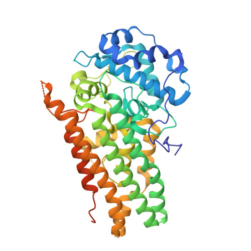
Structural insights into substrate and inhibitor binding sites in human indoleamine 2,3-dioxygenase 1.

Lewis-Ballester, A.Pham, K.N.Batabyal, D.Karkashon, S.Bonanno, J.B.Poulos, T.L.Yeh, S.R.

(2017) Nat Commun 8: 1693-1693

  • DOI: https://doi.org/10.1038/s41467-017-01725-8
  • Primary Citation of Related Structures:  
    5WMU, 5WMV, 5WMW, 5WMX, 5WN8

  • PubMed Abstract: 

    Human indoleamine 2,3-dioxygenase 1 (hIDO1) is an attractive cancer immunotherapeutic target owing to its role in promoting tumoral immune escape. However, drug development has been hindered by limited structural information. Here, we report the crystal structures of hIDO1 in complex with its substrate, Trp, an inhibitor, epacadostat, and/or an effector, indole ethanol (IDE). The data reveal structural features of the active site (Sa) critical for substrate activation; in addition, they disclose a new inhibitor-binding mode and a distinct small molecule binding site (Si). Structure-guided mutation of a critical residue, F270, to glycine perturbs the Si site, allowing structural determination of an inhibitory complex, where both the Sa and Si sites are occupied by Trp. The Si site offers a novel target site for allosteric inhibitors and a molecular explanation for the previously baffling substrate-inhibition behavior of the enzyme. Taken together, the data open exciting new avenues for structure-based drug design.


  • Organizational Affiliation

    Department of Physiology and Biophysics, Albert Einstein College of Medicine, Bronx, NY, 10461, USA.


Macromolecules
Find similar proteins by:  (by identity cutoff)  |  3D Structure
Entity ID: 1
MoleculeChains Sequence LengthOrganismDetailsImage
Indoleamine 2,3-dioxygenase 1
A, B
425Homo sapiensMutation(s): 0 
Gene Names: IDO1IDOINDO
EC: 1.13.11.52
UniProt & NIH Common Fund Data Resources
Find proteins for P14902 (Homo sapiens)
Explore P14902 
Go to UniProtKB:  P14902
PHAROS:  P14902
GTEx:  ENSG00000131203 
Entity Groups  
Sequence Clusters30% Identity50% Identity70% Identity90% Identity95% Identity100% Identity
UniProt GroupP14902
Sequence Annotations
Expand
  • Reference Sequence
Experimental Data & Validation

Experimental Data

  • Method: X-RAY DIFFRACTION
  • Resolution: 2.40 Å
  • R-Value Free: 0.258 
  • R-Value Work: 0.214 
  • R-Value Observed: 0.216 
  • Space Group: P 21 21 21
Unit Cell:
Length ( Å )Angle ( ˚ )
a = 89.301α = 90
b = 97.768β = 90
c = 125.643γ = 90
Software Package:
Software NamePurpose
REFMACrefinement
XDSdata reduction
Aimlessdata scaling
PHASERphasing

Structure Validation

View Full Validation Report


Ligand Structure Quality Assessment 


Entry History & Funding Information

Deposition Data


Funding OrganizationLocationGrant Number
National Institutes of Health/National Cancer Institute (NIH/NCI)United StatesGM115773
National Science Foundation (NSF, United States)United StatesCHE-1404929
National Institutes of Health/National Center for Research Resources (NIH/NCRR)United StatesGM57353

Revision History  (Full details and data files)

  • Version 1.0: 2017-12-06
    Type: Initial release
  • Version 1.1: 2019-11-27
    Changes: Author supporting evidence
  • Version 1.2: 2023-10-04
    Changes: Data collection, Database references, Derived calculations, Refinement description