5WII | pdb_00005wii

TraE protein in complex with 2-Chloroisonicotinic Acid


Experimental Data Snapshot

  • Method: X-RAY DIFFRACTION
  • Resolution: 2.79 Å
  • R-Value Free: 
    0.357 (Depositor), 0.356 (DCC) 
  • R-Value Work: 
    0.298 (Depositor), 0.309 (DCC) 
  • R-Value Observed: 
    0.300 (Depositor) 

Starting Model: experimental
View more details

wwPDB Validation 3D Report Full Report

Validation slider image for 5WII

Ligand Structure Quality Assessment 


This is version 1.1 of the entry. See complete history

Literature

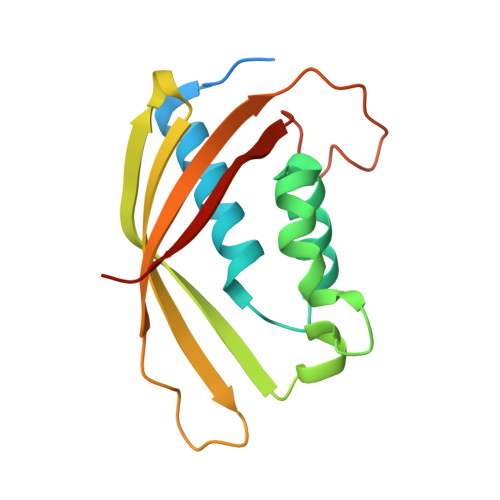
Fragment-based screening identifies novel targets for inhibitors of conjugative transfer of antimicrobial resistance by plasmid pKM101.

Casu, B.Arya, T.Bessette, B.Baron, C.

(2017) Sci Rep 7: 14907-14907

  • DOI: https://doi.org/10.1038/s41598-017-14953-1
  • Primary Citation Related Structures: 
    5WIC, 5WII, 5WIO, 5WIP

  • PubMed Abstract: 

    The increasing frequency of antimicrobial resistance is a problem of global importance. Novel strategies are urgently needed to understand and inhibit antimicrobial resistance gene transmission that is mechanistically related to bacterial virulence functions. The conjugative transfer of plasmids by type IV secretion systems is a major contributor to antimicrobial resistance gene transfer. Here, we present a structure-based strategy to identify inhibitors of type IV secretion system-mediated bacterial conjugation. Using differential scanning fluorimetry we screened a fragment library and identified molecules that bind the essential TraE protein of the plasmid pKM101 conjugation machinery. Co-crystallization revealed that fragments bind two alternative sites of the protein and one of them is a novel inhibitor binding site. Based on the structural information on fragment binding we designed novel small molecules that have improved binding affinity. These molecules inhibit the dimerization of TraE, bind to both inhibitor binding sites on TraE and inhibit the conjugative transfer of plasmid pKM101. The strategy presented here is generally applicable for the structure-based design of inhibitors of antimicrobial resistance gene transfer and of bacterial virulence.


  • Organizational Affiliation
    • Department of Biochemistry and Molecular Medicine, Faculty of Medicine, Université de Montréal, 2900 Boulevard Édouard-Montpetit, Montreal, QC, H3T 1J4, Canada.

Macromolecule Content 

  • Total Structure Weight: 75.43 kDa 
  • Atom Count: 4,514 
  • Modeled Residue Count: 560 
  • Deposited Residue Count: 652 
  • Unique protein chains: 1

Macromolecules

Find similar proteins by:|  3D Structure
Entity ID: 1
MoleculeChains  Sequence LengthOrganismDetailsImage
Conjugal transfer protein
A, B, C, D
163Escherichia coliMutation(s): 0 
Gene Names: 
UniProt
Find proteins for Q17U16 (Escherichia coli)
Explore Q17U16 
Go to UniProtKB:  Q17U16
Entity Groups
Sequence Clusters30% Identity50% Identity70% Identity90% Identity95% Identity100% Identity
UniProt GroupQ17U16
Sequence Annotations
Expand
Reference Sequence

Small Molecules

Ligands 1 Unique
IDChains Name / Formula / InChI Key2D Diagram3D Interactions
AO4

Query on AO4


Download:Ideal Coordinates CCD File
E [auth A]2-chloropyridine-4-carboxylic acid
C6 H4 Cl N O2
QXCOHSRHFCHCHN-UHFFFAOYSA-N

Experimental Data & Validation

Experimental Data

  • Method: X-RAY DIFFRACTION
  • Resolution: 2.79 Å
  • R-Value Free:  0.357 (Depositor), 0.356 (DCC) 
  • R-Value Work:  0.298 (Depositor), 0.309 (DCC) 
  • R-Value Observed: 0.300 (Depositor) 
Space Group: C 2 2 21
Unit Cell:
Length ( Å )Angle ( ˚ )
a = 111.799α = 90
b = 124.116β = 90
c = 109.993γ = 90
Software Package:
Software NamePurpose
PHENIXrefinement
HKL-2000data reduction
HKL-2000data scaling
PHENIXphasing

Structure Validation

View Full Validation Report


Ligand Structure Quality Assessment 


Entry History 

Deposition Data

Revision History  (Full details and data files)

  • Version 1.0: 2017-11-15
    Type: Initial release
  • Version 1.1: 2023-10-04
    Changes: Data collection, Database references, Refinement description