5WGB | pdb_00005wgb

Crystal Structure of the Human mitochondrial Cysteine Desulfurase in complex with ISD11 and E. coli ACP1 protein at 2.75A


Experimental Data Snapshot

  • Method: X-RAY DIFFRACTION
  • Resolution: 2.75 Å
  • R-Value Free: 
    0.255 (Depositor), 0.257 (DCC) 
  • R-Value Work: 
    0.213 (Depositor), 0.217 (DCC) 
  • R-Value Observed: 
    0.216 (Depositor) 

Starting Model: experimental
View more details

wwPDB Validation 3D Report Full Report

Validation slider image for 5WGB

Ligand Structure Quality Assessment 


This is version 1.2 of the entry. See complete history

Literature

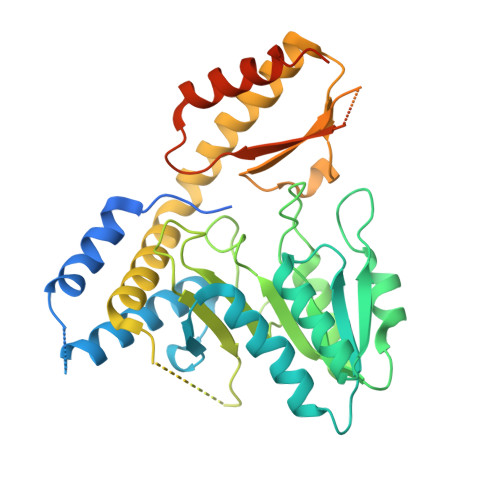
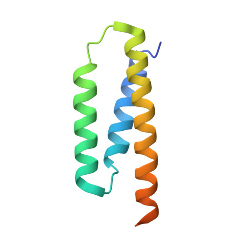
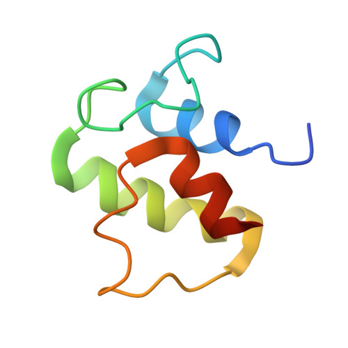
Structure and functional dynamics of the mitochondrial Fe/S cluster synthesis complex.

Boniecki, M.T.Freibert, S.A.Muhlenhoff, U.Lill, R.Cygler, M.

(2017) Nat Commun 8: 1287-1287

  • DOI: https://doi.org/10.1038/s41467-017-01497-1
  • Primary Citation Related Structures: 
    5WGB, 5WKP, 5WLW

  • PubMed Abstract: 

    Iron-sulfur (Fe/S) clusters are essential protein cofactors crucial for many cellular functions including DNA maintenance, protein translation, and energy conversion. De novo Fe/S cluster synthesis occurs on the mitochondrial scaffold protein ISCU and requires cysteine desulfurase NFS1, ferredoxin, frataxin, and the small factors ISD11 and ACP (acyl carrier protein). Both the mechanism of Fe/S cluster synthesis and function of ISD11-ACP are poorly understood. Here, we present crystal structures of three different NFS1-ISD11-ACP complexes with and without ISCU, and we use SAXS analyses to define the 3D architecture of the complete mitochondrial Fe/S cluster biosynthetic complex. Our structural and biochemical studies provide mechanistic insights into Fe/S cluster synthesis at the catalytic center defined by the active-site Cys of NFS1 and conserved Cys, Asp, and His residues of ISCU. We assign specific regulatory rather than catalytic roles to ISD11-ACP that link Fe/S cluster synthesis with mitochondrial lipid synthesis and cellular energy status.


  • Organizational Affiliation
    • Department of Biochemistry, University of Saskatchewan, 107 Wiggins Road, Saskatoon, SK, Canada, S7N 5E5.

Macromolecule Content 

  • Total Structure Weight: 67.46 kDa 
  • Atom Count: 3,247 
  • Modeled Residue Count: 437 
  • Deposited Residue Count: 594 
  • Unique protein chains: 3

Macromolecules

Find similar proteins by:|  3D Structure
Entity ID: 1
MoleculeChains  Sequence LengthOrganismDetailsImage
Cysteine desulfurase, mitochondrial426Homo sapiensMutation(s): 0 
Gene Names: NFS1NIFSHUSSY-08
EC: 2.8.1.7
UniProt & NIH Common Fund Data Resources
Find proteins for Q9Y697 (Homo sapiens)
Explore Q9Y697 
Go to UniProtKB:  Q9Y697
PHAROS:  Q9Y697
GTEx:  ENSG00000244005 
Entity Groups
Sequence Clusters30% Identity50% Identity70% Identity90% Identity95% Identity100% Identity
UniProt GroupQ9Y697
Sequence Annotations
Expand
Reference Sequence
Find similar proteins by:|  3D Structure
Entity ID: 2
MoleculeChains  Sequence LengthOrganismDetailsImage
LYR motif-containing protein 491Homo sapiensMutation(s): 0 
Gene Names: LYRM4C6orf149ISD11CGI-203
UniProt & NIH Common Fund Data Resources
Find proteins for Q9HD34 (Homo sapiens)
Explore Q9HD34 
Go to UniProtKB:  Q9HD34
PHAROS:  Q9HD34
GTEx:  ENSG00000214113 
Entity Groups
Sequence Clusters30% Identity50% Identity70% Identity90% Identity95% Identity100% Identity
UniProt GroupQ9HD34
Sequence Annotations
Expand
Reference Sequence
Find similar proteins by:|  3D Structure
Entity ID: 3
MoleculeChains  Sequence LengthOrganismDetailsImage
Acyl carrier protein77Escherichia coli S88Mutation(s): 0 
Gene Names: acpPECS88_1108
UniProt
Find proteins for B7MJ81 (Escherichia coli O45:K1 (strain S88 / ExPEC))
Explore B7MJ81 
Go to UniProtKB:  B7MJ81
Entity Groups
Sequence Clusters30% Identity50% Identity70% Identity90% Identity95% Identity100% Identity
UniProt GroupB7MJ81
Sequence Annotations
Expand
Reference Sequence

Small Molecules

Ligands 2 Unique
IDChains Name / Formula / InChI Key2D Diagram3D Interactions
8Q1

Query on 8Q1


Download:Ideal Coordinates CCD File
E [auth C]S-[2-({N-[(2R)-2-hydroxy-3,3-dimethyl-4-(phosphonooxy)butanoyl]-beta-alanyl}amino)ethyl] dodecanethioate
C23 H45 N2 O8 P S
MVHUOSAYFQKAMT-NRFANRHFSA-N
PLP

Query on PLP


Download:Ideal Coordinates CCD File
D [auth A]PYRIDOXAL-5'-PHOSPHATE
C8 H10 N O6 P
NGVDGCNFYWLIFO-UHFFFAOYSA-N

Experimental Data & Validation

Experimental Data

  • Method: X-RAY DIFFRACTION
  • Resolution: 2.75 Å
  • R-Value Free:  0.255 (Depositor), 0.257 (DCC) 
  • R-Value Work:  0.213 (Depositor), 0.217 (DCC) 
  • R-Value Observed: 0.216 (Depositor) 
Space Group: H 3 2
Unit Cell:
Length ( Å )Angle ( ˚ )
a = 140.841α = 90
b = 140.841β = 90
c = 203.301γ = 120
Software Package:
Software NamePurpose
PHENIXrefinement
XDSdata reduction
XDSdata scaling
PHASERphasing

Structure Validation

View Full Validation Report


Ligand Structure Quality Assessment 


Entry History 

Deposition Data

Revision History  (Full details and data files)

  • Version 1.0: 2017-07-26
    Type: Initial release
  • Version 1.1: 2017-11-15
    Changes: Database references, Refinement description, Source and taxonomy
  • Version 1.2: 2023-10-04
    Changes: Data collection, Database references, Refinement description