5W38

1.80A resolution structure of human IgG3 Fc (N392K)


Experimental Data Snapshot

  • Method: X-RAY DIFFRACTION
  • Resolution: 1.80 Å
  • R-Value Free: 0.223 
  • R-Value Work: 0.183 
  • R-Value Observed: 0.186 

wwPDB Validation   3D Report Full Report


This is version 2.1 of the entry. See complete history


Literature

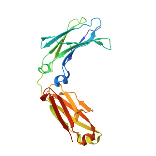
Structural characterization of the Man5 glycoform of human IgG3 Fc.

Shah, I.S.Lovell, S.Mehzabeen, N.Battaile, K.P.Tolbert, T.J.

(2017) Mol Immunol 92: 28-37

  • DOI: https://doi.org/10.1016/j.molimm.2017.10.001
  • Primary Citation of Related Structures:  
    5W38

  • PubMed Abstract: 

    Immunoglobulin G (IgG) consists of four subclasses in humans: IgG1, IgG2, IgG3 and IgG4, which are highly conserved but have unique differences that result in subclass-specific effector functions. Though IgG1 is the most extensively studied IgG subclass, study of other subclasses is important to understand overall immune function and for development of new therapeutics. When compared to IgG1, IgG3 exhibits a similar binding profile to Fcγ receptors and stronger activation of complement. All IgG subclasses are glycosylated at N297, which is required for Fcγ receptor and C1q complement binding as well as maintaining optimal Fc conformation. We have determined the crystal structure of homogenously glycosylated human IgG3 Fc with a GlcNAc 2 Man 5 (Man5) high mannose glycoform at 1.8Å resolution and compared its structural features with published structures from the other IgG subclasses. Although the overall structure of IgG3 Fc is similar to that of other subclasses, some structural perturbations based on sequence differences were revealed. For instance, the presence of R435 in IgG3 (and H435 in the other IgG subclasses) has been implicated to result in IgG3-specific properties related to binding to protein A, protein G and the neonatal Fc receptor (FcRn). The IgG3 Fc structure helps to explain some of these differences. Additionally, protein-glycan contacts observed in the crystal structure appear to correlate with IgG3 affinity for Fcγ receptors as shown by binding studies with IgG3 Fc glycoforms. Finally, this IgG3 Fc structure provides a template for further studies aimed at engineering the Fc for specific gain of function.


  • Organizational Affiliation

    Department of Pharmaceutical Chemistry, University of Kansas, Lawrence, KS, USA.


Macromolecules
Find similar proteins by:  (by identity cutoff)  |  3D Structure
Entity ID: 1
MoleculeChains Sequence LengthOrganismDetailsImage
Immunoglobulin heavy constant gamma 3
A, B
223Homo sapiensMutation(s): 1 
Gene Names: IGHG3
UniProt & NIH Common Fund Data Resources
Find proteins for P01860 (Homo sapiens)
Explore P01860 
Go to UniProtKB:  P01860
PHAROS:  P01860
Entity Groups  
Sequence Clusters30% Identity50% Identity70% Identity90% Identity95% Identity100% Identity
UniProt GroupP01860
Sequence Annotations
Expand
  • Reference Sequence
Oligosaccharides

Help

Entity ID: 2
MoleculeChains Length2D Diagram Glycosylation3D Interactions
beta-D-mannopyranose-(1-4)-2-acetamido-2-deoxy-beta-D-glucopyranose-(1-4)-2-acetamido-2-deoxy-beta-D-glucopyranose
C, D
3N-Glycosylation
Glycosylation Resources
GlyTouCan:  G15407YE
GlyCosmos:  G15407YE
GlyGen:  G15407YE
Small Molecules
Ligands 1 Unique
IDChains Name / Formula / InChI Key2D Diagram3D Interactions
FLC
Query on FLC

Download Ideal Coordinates CCD File 
E [auth A]CITRATE ANION
C6 H5 O7
KRKNYBCHXYNGOX-UHFFFAOYSA-K
Experimental Data & Validation

Experimental Data

  • Method: X-RAY DIFFRACTION
  • Resolution: 1.80 Å
  • R-Value Free: 0.223 
  • R-Value Work: 0.183 
  • R-Value Observed: 0.186 
  • Space Group: P 1 21 1
Unit Cell:
Length ( Å )Angle ( ˚ )
a = 64.429α = 90
b = 60.443β = 109.24
c = 71.396γ = 90
Software Package:
Software NamePurpose
Aimlessdata scaling
PHASERphasing
PHENIXrefinement
PDB_EXTRACTdata extraction
XDSdata reduction

Structure Validation

View Full Validation Report


Entry History 

Deposition Data

Revision History  (Full details and data files)

  • Version 1.0: 2017-10-18
    Type: Initial release
  • Version 1.1: 2017-11-01
    Changes: Database references
  • Version 2.0: 2020-07-29
    Type: Remediation
    Reason: Carbohydrate remediation
    Changes: Atomic model, Data collection, Derived calculations, Structure summary
  • Version 2.1: 2023-10-04
    Changes: Data collection, Database references, Refinement description, Structure summary