5VZU | pdb_00005vzu

Crystal structure of the Skp1-FBXO31-cyclin D1 complex

  • Classification: CELL CYCLE
  • Organism(s): Homo sapiens
  • Expression System: Escherichia coli BL21(DE3)
  • Mutation(s): Yes 

  • Deposited: 2017-05-29 Released: 2018-01-17 
  • Deposition Author(s): Li, Y., Jin, K., Hao, B.
  • Funding Organization(s): National Institutes of Health/National Institute of General Medical Sciences (NIH/NIGMS), Connecticut Regenerative Medicine Research Fund

Experimental Data Snapshot

  • Method: X-RAY DIFFRACTION
  • Resolution: 2.70 Å
  • R-Value Free: 
    0.233 (Depositor), 0.236 (DCC) 
  • R-Value Work: 
    0.180 (Depositor), 0.184 (DCC) 
  • R-Value Observed: 
    0.183 (Depositor) 

Starting Model: experimental
View more details

wwPDB Validation 3D Report Full Report

Validation slider image for 5VZU

This is version 1.3 of the entry. See complete history

Literature

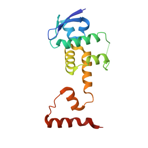
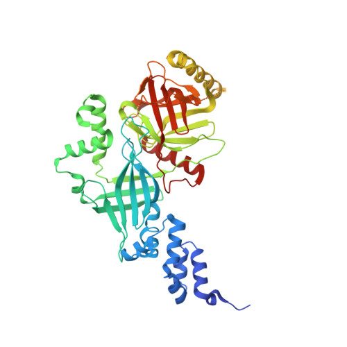
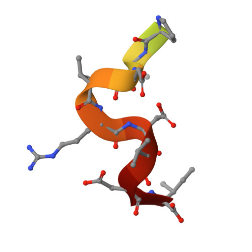
Structural basis of the phosphorylation-independent recognition of cyclin D1 by the SCFFBXO31 ubiquitin ligase.

Li, Y.Jin, K.Bunker, E.Zhang, X.Luo, X.Liu, X.Hao, B.

(2018) Proc Natl Acad Sci U S A 115: 319-324

  • DOI: https://doi.org/10.1073/pnas.1708677115
  • Primary Citation Related Structures: 
    5VZT, 5VZU

  • PubMed Abstract: 

    Ubiquitin-dependent proteolysis of cyclin D1 is associated with normal and tumor cell proliferation and survival. The SCF FBXO31 (Skp1-Cul1-Rbx1-FBXO31) ubiquitin ligase complex mediates genotoxic stress-induced cyclin D1 degradation. Previous studies have suggested that cyclin D1 levels are maintained at steady state by phosphorylation-dependent nuclear export and subsequent proteolysis in the cytoplasm. Here we present the crystal structures of the Skp1-FBXO31 complex alone and bound to a phosphorylated cyclin D1 C-terminal peptide. FBXO31 possesses a unique substrate-binding domain consisting of two β-barrel motifs, whereas cyclin D1 binds to FBXO31 by tucking its free C-terminal carboxylate tail into an open cavity of the C-terminal FBXO31 β-barrel. Biophysical and functional studies demonstrate that SCF FBXO31 is capable of recruiting and ubiquitinating cyclin D1 in a phosphorylation-independent manner. Our findings provide a conceptual framework for understanding the substrate specificity of the F-box protein FBXO31 and the mechanism of FBXO31-regulated cyclin D1 protein turnover.


  • Organizational Affiliation
    • Department of Molecular Biology and Biophysics, University of Connecticut Health Center, Farmington, CT 06030.

Macromolecule Content 

  • Total Structure Weight: 150.14 kDa 
  • Atom Count: 9,361 
  • Modeled Residue Count: 1,138 
  • Deposited Residue Count: 1,308 
  • Unique protein chains: 3

Macromolecules

Find similar proteins by:|  3D Structure
Entity ID: 1
MoleculeChains  Sequence LengthOrganismDetailsImage
S-phase kinase-associated protein 1
A, C
149Homo sapiensMutation(s): 1 
Gene Names: SKP1EMC19OCP2SKP1ATCEB1L
UniProt & NIH Common Fund Data Resources
Find proteins for P63208 (Homo sapiens)
Explore P63208 
Go to UniProtKB:  P63208
PHAROS:  P63208
GTEx:  ENSG00000113558 
Entity Groups
Sequence Clusters30% Identity50% Identity70% Identity90% Identity95% Identity100% Identity
UniProt GroupP63208
Sequence Annotations
Expand
Reference Sequence
Find similar proteins by:|  3D Structure
Entity ID: 2
MoleculeChains  Sequence LengthOrganismDetailsImage
F-box only protein 31
B, D
488Homo sapiensMutation(s): 0 
Gene Names: FBXO31FBX14FBX31PP2386
UniProt & NIH Common Fund Data Resources
Find proteins for Q5XUX0 (Homo sapiens)
Explore Q5XUX0 
Go to UniProtKB:  Q5XUX0
PHAROS:  Q5XUX0
GTEx:  ENSG00000103264 
Entity Groups
Sequence Clusters30% Identity50% Identity70% Identity90% Identity95% Identity100% Identity
UniProt GroupQ5XUX0
Sequence Annotations
Expand
Reference Sequence
Find similar proteins by:  Sequence   |   3D Structure  
Entity ID: 3
MoleculeChains  Sequence LengthOrganismDetailsImage
Cyclin D1
E, F
17Homo sapiensMutation(s): 0 
UniProt & NIH Common Fund Data Resources
Find proteins for P24385 (Homo sapiens)
Explore P24385 
Go to UniProtKB:  P24385
PHAROS:  P24385
GTEx:  ENSG00000110092 
Entity Groups
Sequence Clusters30% Identity50% Identity70% Identity90% Identity95% Identity100% Identity
UniProt GroupP24385
Sequence Annotations
Expand
Reference Sequence

Small Molecules

Ligands 2 Unique
IDChains Name / Formula / InChI Key2D Diagram3D Interactions
PO4

Query on PO4


Download:Ideal Coordinates CCD File
H [auth B]
I [auth B]
J [auth B]
K [auth B]
L [auth B]
H [auth B],
I [auth B],
J [auth B],
K [auth B],
L [auth B],
N [auth D],
O [auth D],
P [auth D],
Q [auth D],
R [auth D]
PHOSPHATE ION
O4 P
NBIIXXVUZAFLBC-UHFFFAOYSA-K
ZN

Query on ZN


Download:Ideal Coordinates CCD File
G [auth B],
M [auth D]
ZINC ION
Zn
PTFCDOFLOPIGGS-UHFFFAOYSA-N
Modified Residues  1 Unique
IDChains TypeFormula2D DiagramParent
TPO
Query on TPO
E, F
L-PEPTIDE LINKINGC4 H10 N O6 PTHR

Experimental Data & Validation

Experimental Data

  • Method: X-RAY DIFFRACTION
  • Resolution: 2.70 Å
  • R-Value Free:  0.233 (Depositor), 0.236 (DCC) 
  • R-Value Work:  0.180 (Depositor), 0.184 (DCC) 
  • R-Value Observed: 0.183 (Depositor) 
Space Group: C 1 2 1
Unit Cell:
Length ( Å )Angle ( ˚ )
a = 87.679α = 90
b = 156.235β = 104.21
c = 155.522γ = 90
Software Package:
Software NamePurpose
REFMACrefinement
HKL-2000data scaling
PDB_EXTRACTdata extraction
HKL-2000data reduction
PHASERphasing

Structure Validation

View Full Validation Report


Entry History 

& Funding Information

Deposition Data


Funding OrganizationLocationGrant Number
National Institutes of Health/National Institute of General Medical Sciences (NIH/NIGMS)United StatesGM099948
Connecticut Regenerative Medicine Research FundUnited States15-RMA-UCHC-04

Revision History  (Full details and data files)

  • Version 1.0: 2018-01-17
    Type: Initial release
  • Version 1.1: 2020-01-01
    Changes: Author supporting evidence
  • Version 1.2: 2023-10-04
    Changes: Data collection, Database references, Refinement description
  • Version 1.3: 2024-11-20
    Changes: Structure summary