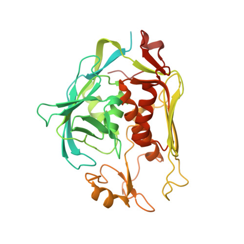
Crystal structure of UDP-3-O-[3-hydroxymyristoyl] N-acetylglucosamine deacetylase (LpxC) from Pseudomonas aeruginosa in complex with CHIR-090 inhibitor
Delker, S.L., Mayclin, S.J., Phan, J.N., Abendroth, J., Lorimer, D., Edwards, T.E.To be published.