Structure and Misfolding of the Flexible Tripartite Coiled-Coil Domain of Glaucoma-Associated Myocilin.
Hill, S.E., Nguyen, E., Donegan, R.K., Patterson-Orazem, A.C., Hazel, A., Gumbart, J.C., Lieberman, R.L.(2017) Structure 25: 1697-1707.e5
- PubMed: 29056483 Search on PubMedSearch on PubMed Central
- DOI: https://doi.org/10.1016/j.str.2017.09.008
- Primary Citation Related Structures:
5VR2 - PubMed Abstract:
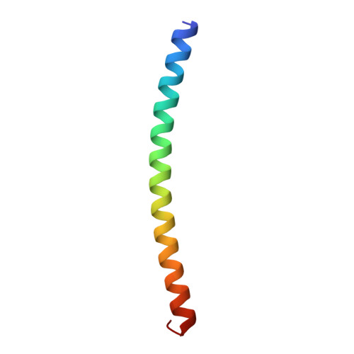
Glaucoma-associated myocilin is a member of the olfactomedins, a protein family involved in neuronal development and human diseases. Molecular studies of the myocilin N-terminal coiled coil demonstrate a unique tripartite architecture: a Y-shaped parallel dimer-of-dimers with distinct tetramer and dimer regions. The structure of the dimeric C-terminal 7-heptad repeats elucidates an unexpected repeat pattern involving inter-strand stabilization by oppositely charged residues. Molecular dynamics simulations reveal an alternate accessible conformation in which the terminal inter-strand disulfide limits the extent of unfolding and results in a kinked configuration. By inference, full-length myocilin is also branched, with two pairs of C-terminal olfactomedin domains. Selected variants within the N-terminal region alter the apparent quaternary structure of myocilin but do so without compromising stability or causing aggregation. In addition to increasing our structural knowledge of naturally occurring extracellular coiled coils and biomedically important olfactomedins, this work broadens the scope of protein misfolding in the pathogenesis of myocilin-associated glaucoma.
- School of Chemistry and Biochemistry, Georgia Institute of Technology, Atlanta, GA 30332, USA.
Organizational Affiliation: