


X-ray co-structure of nuclear receptor ROR-gammat Ligand Binding Domain with a inverse agonist and SRC2 peptide
Li, X.To be published.
Experimental Data Snapshot
Entity ID: 1 | |||||
|---|---|---|---|---|---|
| Molecule | Chains | Sequence Length | Organism | Details | Image |
| Nuclear receptor ROR-gamma, SRC2 chimera | 280 | Homo sapiens | Mutation(s): 0 Gene Names: RORC, NR1F3, RORG, RZRG | 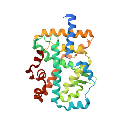 | |
UniProt & NIH Common Fund Data Resources | |||||
GTEx: ENSG00000143365 | |||||
GTEx: ENSG00000140396 | |||||
Entity Groups | |||||
| Sequence Clusters | 30% Identity50% Identity70% Identity90% Identity95% Identity100% Identity | ||||
| UniProt Groups | P51449Q15596 | ||||
Sequence AnnotationsExpand | |||||
Reference Sequence | |||||
| Ligands 2 Unique | |||||
|---|---|---|---|---|---|
| ID | Chains | Name / Formula / InChI Key | 2D Diagram | 3D Interactions | |
| 9GS Download:Ideal Coordinates CCD File | C [auth A] | 5'-(4-cyclopropyl-6-methoxypyrimidin-5-yl)-N-{[4-(ethylsulfonyl)phenyl]methyl}spiro[cyclopentane-1,3'-pyrrolo[3,2-b]pyridine]-1'(2'H)-carboxamide C29 H33 N5 O4 S PWIXSNKHPMGRRW-UHFFFAOYSA-N |  | ||
| NA Download:Ideal Coordinates CCD File | B [auth A] | SODIUM ION Na FKNQFGJONOIPTF-UHFFFAOYSA-N |  | ||
| Length ( Å ) | Angle ( ˚ ) |
|---|---|
| a = 61.325 | α = 90 |
| b = 61.325 | β = 90 |
| c = 155.578 | γ = 90 |
| Software Name | Purpose |
|---|---|
| BUSTER | refinement |
| d*TREK | data reduction |
| d*TREK | data scaling |
| PHENIX | phasing |


