Reaction mechanism of the bioluminescent protein mnemiopsin1 revealed by X-ray crystallography and QM/MM simulations.
Molakarimi, M., Gorman, M.A., Mohseni, A., Pashandi, Z., Taghdir, M., Naderi-Manesh, H., Sajedi, R.H., Parker, M.W.(2019) J Biological Chem 294: 20-27
- PubMed: 30420427 Search on PubMedSearch on PubMed Central
- DOI: https://doi.org/10.1074/jbc.RA118.006053
- Primary Citation Related Structures:
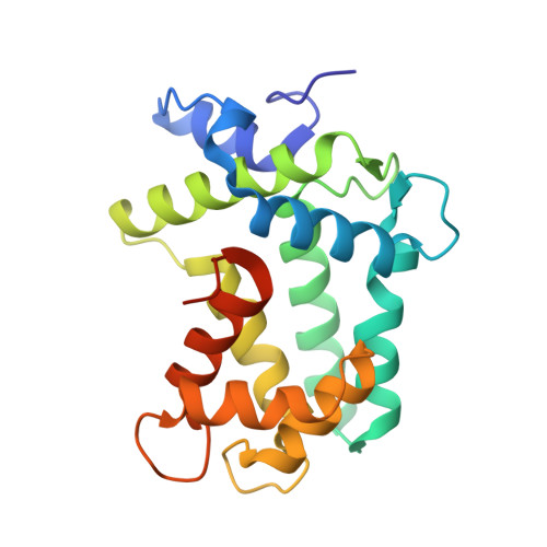
5VP3 - PubMed Abstract:
Bioluminescence of a variety of marine organisms, mostly cnidarians and ctenophores, is carried out by Ca 2+ -dependent photoproteins. The mechanism of light emission operates via the same reaction in both animal families. Despite numerous studies on the ctenophore photoprotein family, the detailed catalytic mechanism and arrangement of amino acid residues surrounding the chromophore in this family are a mystery. Here, we report the crystal structure of Cd 2+ -loaded apo-mnemiopsin1, a member of the ctenophore family, at 2.15 Å resolution and used quantum mechanics/molecular mechanics (QM/MM) to investigate its reaction mechanism. The simulations suggested that an Asp-156-Arg-39-Tyr-202 triad creates a hydrogen-bonded network to facilitate the transfer of a proton from the 2-hydroperoxy group of the chromophore coelenterazine to bulk solvent. We identified a water molecule in the coelenteramide-binding cavity that forms a hydrogen bond with the amide nitrogen atom of coelenteramide, which, in turn, is hydrogen-bonded via another water molecule to Tyr-131. This observation supports the hypothesis that the function of the coelenteramide-bound water molecule is to catalyze the 2-hydroperoxycoelenterazine decarboxylation reaction by protonation of a dioxetanone anion, thereby triggering the bioluminescence reaction in the ctenophore photoprotein family.
- Department of Biochemistry, Faculty of Biological Sciences, Tarbiat Modares University, Tehran 14115-154, Iran.
Organizational Affiliation: