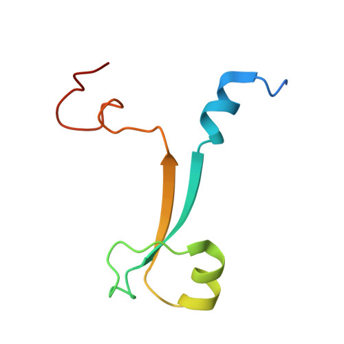
5VMD

Crystal structure of UBR-box from UBR6 in a domain-swapping conformation


Experimental Data Snapshot

  • Method: X-RAY DIFFRACTION
  • Resolution: 2.20 Å
  • R-Value Free: 0.247 
  • R-Value Work: 0.195 
  • R-Value Observed: 0.197 

wwPDB Validation   3D Report Full Report


This is version 1.4 of the entry. See complete history


Literature

Crystal structure of the UBR-box from UBR6/FBXO11 reveals domain swapping mediated by zinc binding.

Munoz-Escobar, J.Kozlov, G.Gehring, K.

(2017) Protein Sci 26: 2092-2097

  • DOI: https://doi.org/10.1002/pro.3227
  • Primary Citation of Related Structures:  
    5VMD

  • PubMed Abstract: 
  • The UBR-box is a 70-residue zinc finger domain present in the UBR family of E3 ubiquitin ligases that directly binds N-terminal degradation signals in substrate proteins. UBR6, also called FBXO11, is an UBR-box containing E3 ubiquitin ligase that does not bind N-terminal signals ...

    The UBR-box is a 70-residue zinc finger domain present in the UBR family of E3 ubiquitin ligases that directly binds N-terminal degradation signals in substrate proteins. UBR6, also called FBXO11, is an UBR-box containing E3 ubiquitin ligase that does not bind N-terminal signals. Here, we present the crystal structure of the UBR-box domain from human UBR6. The dimeric crystal structure reveals a unique form of domain swapping mediated by zinc coordination, where three independent protein chains come together to regenerate the topology of the monomeric UBR-box fold. Analysis of the structure suggests that the absence of N-terminal residue binding arises from the lack of an amino acid binding pocket.


    Organizational Affiliation

    Department of Biochemistry, Groupe de Recherche Axé sur la Structure des Protéines, McGill University, Montreal, Quebec, H3G0B1, Canada.


Macromolecules
Find similar proteins by:  (by identity cutoff)  |  3D Structure
Entity ID: 1
MoleculeChainsSequence LengthOrganismDetailsImage
F-box only protein 11A, B, C, D77Homo sapiensMutation(s): 0 
Gene Names: FBXO11FBX11PRMT9VIT1UG063H01
UniProt & NIH Common Fund Data Resources
Find proteins for Q86XK2 (Homo sapiens)
Explore Q86XK2 
Go to UniProtKB:  Q86XK2
PHAROS:  Q86XK2
Entity Groups  
Sequence Clusters30% Identity50% Identity70% Identity90% Identity95% Identity100% Identity
UniProt GroupQ86XK2
Protein Feature View
Expand
  • Reference Sequence
Small Molecules
Ligands 2 Unique
IDChainsName / Formula / InChI Key2D Diagram3D Interactions
ZN
Query on ZN

Download Ideal Coordinates CCD File 
E [auth A],
F [auth A],
G [auth A],
H [auth A],
I [auth A],
E [auth A],
F [auth A],
G [auth A],
H [auth A],
I [auth A],
N [auth B],
O [auth B],
P [auth B],
Q [auth B],
R [auth B],
U [auth C],
V [auth C],
X [auth D],
Y [auth D]
ZINC ION
Zn
PTFCDOFLOPIGGS-UHFFFAOYSA-N
 Ligand Interaction
EDO
Query on EDO

Download Ideal Coordinates CCD File 
AA [auth D],
J [auth A],
K [auth A],
L [auth A],
M [auth A],
AA [auth D],
J [auth A],
K [auth A],
L [auth A],
M [auth A],
S [auth B],
T [auth B],
W [auth C],
Z [auth D]
1,2-ETHANEDIOL
C2 H6 O2
LYCAIKOWRPUZTN-UHFFFAOYSA-N
 Ligand Interaction
Experimental Data & Validation

Experimental Data

  • Method: X-RAY DIFFRACTION
  • Resolution: 2.20 Å
  • R-Value Free: 0.247 
  • R-Value Work: 0.195 
  • R-Value Observed: 0.197 
  • Space Group: P 21 21 21
Unit Cell:
Length ( Å )Angle ( ˚ )
a = 67.57α = 90
b = 69.2β = 90
c = 82.59γ = 90
Software Package:
Software NamePurpose
PHENIXrefinement
HKL-2000data reduction
HKL-2000data scaling
PHENIXphasing

Structure Validation

View Full Validation Report


Entry History & Funding Information

Deposition Data


Funding OrganizationLocationGrant Number
Canadian Institutes of Health Research (CIHR)Canada--

Revision History  (Full details and data files)

  • Version 1.0: 2017-07-12
    Type: Initial release
  • Version 1.1: 2017-07-26
    Changes: Author supporting evidence, Database references
  • Version 1.2: 2017-08-09
    Changes: Database references
  • Version 1.3: 2017-10-04
    Changes: Database references
  • Version 1.4: 2020-01-08
    Changes: Author supporting evidence