5VE5

Crystal structure of persulfide dioxygenase rhodanese fusion protein with rhodanese domain inactivating mutation (C314S) from Burkholderia phytofirmans in complex with glutathione


Experimental Data Snapshot

  • Method: X-RAY DIFFRACTION
  • Resolution: 2.35 Å
  • R-Value Free: 0.252 
  • R-Value Work: 0.197 
  • R-Value Observed: 0.199 

wwPDB Validation   3D Report Full Report


Ligand Structure Quality Assessment 


This is version 1.3 of the entry. See complete history


Literature

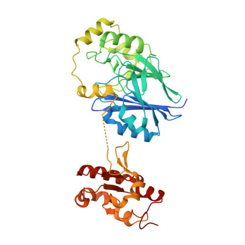
Structural and biochemical analyses indicate that a bacterial persulfide dioxygenase-rhodanese fusion protein functions in sulfur assimilation.

Motl, N.Skiba, M.A.Kabil, O.Smith, J.L.Banerjee, R.

(2017) J Biol Chem 292: 14026-14038

  • DOI: https://doi.org/10.1074/jbc.M117.790170
  • Primary Citation of Related Structures:  
    5VE3, 5VE4, 5VE5

  • PubMed Abstract: 

    Hydrogen sulfide (H 2 S) is a signaling molecule that is toxic at elevated concentrations. In eukaryotes, it is cleared via a mitochondrial sulfide oxidation pathway, which comprises sulfide quinone oxidoreductase, persulfide dioxygenase (PDO), rhodanese, and sulfite oxidase and converts H 2 S to thiosulfate and sulfate. Natural fusions between the non-heme iron containing PDO and rhodanese, a thiol sulfurtransferase, exist in some bacteria. However, little is known about the role of the PDO-rhodanese fusion (PRF) proteins in sulfur metabolism. Herein, we report the kinetic properties and the crystal structure of a PRF from the Gram-negative endophytic bacterium Burkholderia phytofirmans The crystal structures of wild-type PRF and a sulfurtransferase-inactivated C314S mutant with and without glutathione were determined at 1.8, 2.4, and 2.7 Å resolution, respectively. We found that the two active sites are distant and do not show evidence of direct communication. The B. phytofirmans PRF exhibited robust PDO activity and preferentially catalyzed sulfur transfer in the direction of thiosulfate to sulfite and glutathione persulfide; sulfur transfer in the reverse direction was detectable only under limited turnover conditions. Together with the kinetic data, our bioinformatics analysis reveals that B. phytofirmans PRF is poised to metabolize thiosulfate to sulfite in a sulfur assimilation pathway rather than in sulfide stress response as seen, for example, with the Staphylococcus aureus PRF or sulfide oxidation and disposal as observed with the homologous mammalian proteins.


  • Organizational Affiliation

    From the Department of Biological Chemistry, University of Michigan Medical Center, Ann Arbor, Michigan 48109-0600.


Macromolecules
Find similar proteins by:  (by identity cutoff)  |  3D Structure
Entity ID: 1
MoleculeChains Sequence LengthOrganismDetailsImage
BpPRF
A, B, C
377Paraburkholderia phytofirmans PsJNMutation(s): 1 
Gene Names: Bphyt_4191
EC: 1.13.11.18 (PDB Primary Data), 2.8.1.1 (PDB Primary Data)
UniProt
Find proteins for B2TEQ2 (Paraburkholderia phytofirmans (strain DSM 17436 / LMG 22146 / PsJN))
Explore B2TEQ2 
Go to UniProtKB:  B2TEQ2
Entity Groups  
Sequence Clusters30% Identity50% Identity70% Identity90% Identity95% Identity100% Identity
UniProt GroupB2TEQ2
Sequence Annotations
Expand
  • Reference Sequence
Small Molecules
Ligands 5 Unique
IDChains Name / Formula / InChI Key2D Diagram3D Interactions
GSH
Query on GSH

Download Ideal Coordinates CCD File 
E [auth A],
J [auth B],
M [auth C]
GLUTATHIONE
C10 H17 N3 O6 S
RWSXRVCMGQZWBV-WDSKDSINSA-N
PGE
Query on PGE

Download Ideal Coordinates CCD File 
H [auth A]TRIETHYLENE GLYCOL
C6 H14 O4
ZIBGPFATKBEMQZ-UHFFFAOYSA-N
IMD
Query on IMD

Download Ideal Coordinates CCD File 
G [auth A],
O [auth C]
IMIDAZOLE
C3 H5 N2
RAXXELZNTBOGNW-UHFFFAOYSA-O
FE
Query on FE

Download Ideal Coordinates CCD File 
D [auth A],
I [auth B],
L [auth C]
FE (III) ION
Fe
VTLYFUHAOXGGBS-UHFFFAOYSA-N
CL
Query on CL

Download Ideal Coordinates CCD File 
F [auth A],
K [auth B],
N [auth C]
CHLORIDE ION
Cl
VEXZGXHMUGYJMC-UHFFFAOYSA-M
Experimental Data & Validation

Experimental Data

  • Method: X-RAY DIFFRACTION
  • Resolution: 2.35 Å
  • R-Value Free: 0.252 
  • R-Value Work: 0.197 
  • R-Value Observed: 0.199 
  • Space Group: P 65 2 2
Unit Cell:
Length ( Å )Angle ( ˚ )
a = 83.503α = 90
b = 83.503β = 90
c = 547.609γ = 120
Software Package:
Software NamePurpose
XSCALEdata scaling
PHENIXrefinement
PDB_EXTRACTdata extraction
XDSdata reduction

Structure Validation

View Full Validation Report


Ligand Structure Quality Assessment 


Entry History & Funding Information

Deposition Data


Funding OrganizationLocationGrant Number
National Institutes of Health/National Institute of General Medical Sciences (NIH/NIGMS)United StatesGM112455
National Institutes of Health/National Institute of General Medical Sciences (NIH/NIGMS)United StatesT32GM008353

Revision History  (Full details and data files)

  • Version 1.0: 2017-07-19
    Type: Initial release
  • Version 1.1: 2017-09-06
    Changes: Author supporting evidence, Database references
  • Version 1.2: 2020-01-01
    Changes: Author supporting evidence, Derived calculations
  • Version 1.3: 2023-10-04
    Changes: Data collection, Database references, Derived calculations, Refinement description