5VDI

Crystal Structure of Human Glycine Receptor alpha-3 Mutant N38Q Bound to AM-3607, Glycine, and Ivermectin


Experimental Data Snapshot

  • Method: X-RAY DIFFRACTION
  • Resolution: 3.10 Å
  • R-Value Free: 0.256 
  • R-Value Work: 0.232 
  • R-Value Observed: 0.233 

wwPDB Validation   3D Report Full Report


Ligand Structure Quality Assessment 


This is version 1.2 of the entry. See complete history


Literature

Crystal Structures of Human GlyR alpha 3 Bound to Ivermectin.

Huang, X.Chen, H.Shaffer, P.L.

(2017) Structure 25: 945-950.e2

  • DOI: https://doi.org/10.1016/j.str.2017.04.007
  • Primary Citation of Related Structures:  
    5VDI

  • PubMed Abstract: 

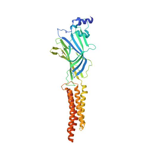
    Ivermectin acts as a positive allosteric modulator of several Cys-loop receptors including the glutamate-gated chloride channels (GluCls), γ-aminobutyric acid receptors (GABA A Rs), glycine receptors (GlyRs), and neuronal α7-nicotinic receptors (α7 nAChRs). The crystal structure of Caenorhabditis elegans GluCl complexed with ivermectin revealed the details of its ivermectin binding site. Although the electron microscopy structure of zebrafish GlyRα1 complexed with ivermectin demonstrated a similar binding orientation, detailed structural information on the ivermectin binding and pore opening for Cys-loop receptors in vertebrates has been elusive. Here we present the crystal structures of human GlyRα3 in complex with ivermectin at 2.85 and 3.08 Å resolution. Our structures allow us to explore in detail the molecular recognition of ivermectin by GlyRs, GABA A Rs, and α7 nAChRs. Comparisons with previous structures reveal how the ivermectin binding expands the ion channel pore. Our results hold promise in structure-based design of GlyR modulators for the treatment of neuropathic pain.


  • Organizational Affiliation

    Department of Molecular Structure and Characterization, Amgen Inc., 360 Binney Street, Cambridge, MA 02142, USA. Electronic address: hxin@amgen.com.


Macromolecules
Find similar proteins by:  (by identity cutoff)  |  3D Structure
Entity ID: 1
MoleculeChains Sequence LengthOrganismDetailsImage
Glycine receptor subunit alpha-3
A, B, C, D, E
362Homo sapiensMutation(s): 1 
Gene Names: GLRA3
Membrane Entity: Yes 
UniProt & NIH Common Fund Data Resources
Find proteins for O75311 (Homo sapiens)
Explore O75311 
Go to UniProtKB:  O75311
PHAROS:  O75311
GTEx:  ENSG00000145451 
Entity Groups  
Sequence Clusters30% Identity50% Identity70% Identity90% Identity95% Identity100% Identity
UniProt GroupO75311
Sequence Annotations
Expand
  • Reference Sequence
Small Molecules
Ligands 4 Unique
IDChains Name / Formula / InChI Key2D Diagram3D Interactions
IVM
Query on IVM

Download Ideal Coordinates CCD File 
H [auth A],
L [auth B],
P [auth C],
T [auth D],
X [auth E]
(2aE,4E,5'S,6S,6'R,7S,8E,11R,13R,15S,17aR,20R,20aR,20bS)-6'-[(2S)-butan-2-yl]-20,20b-dihydroxy-5',6,8,19-tetramethyl-17 -oxo-3',4',5',6,6',10,11,14,15,17,17a,20,20a,20b-tetradecahydro-2H,7H-spiro[11,15-methanofuro[4,3,2-pq][2,6]benzodioxacy clooctadecine-13,2'-pyran]-7-yl 2,6-dideoxy-4-O-(2,6-dideoxy-3-O-methyl-alpha-L-arabino-hexopyranosyl)-3-O-methyl-alpha-L-arabino-hexopyranoside
C48 H74 O14
AZSNMRSAGSSBNP-XPNPUAGNSA-N
7C6
Query on 7C6

Download Ideal Coordinates CCD File 
F [auth A],
J [auth B],
N [auth C],
R [auth D],
V [auth E]
(3S,3aS,9bS)-2-[(2H-1,3-benzodioxol-5-yl)sulfonyl]-3,5-dimethyl-1,2,3,3a,5,9b-hexahydro-4H-pyrrolo[3,4-c][1,6]naphthyridin-4-one
C19 H19 N3 O5 S
MXNQASHPCAMJHG-UCMVZMLTSA-N
GLY
Query on GLY

Download Ideal Coordinates CCD File 
G [auth A],
K [auth B],
O [auth C],
S [auth D],
W [auth E]
GLYCINE
C2 H5 N O2
DHMQDGOQFOQNFH-UHFFFAOYSA-N
ZN
Query on ZN

Download Ideal Coordinates CCD File 
I [auth A],
M [auth B],
Q [auth C],
U [auth D],
Y [auth E]
ZINC ION
Zn
PTFCDOFLOPIGGS-UHFFFAOYSA-N
Binding Affinity Annotations 
IDSourceBinding Affinity
7C6 BindingDB:  5VDI Kd: 11 (nM) from 1 assay(s)
EC50: min: 32, max: 50 (nM) from 2 assay(s)
Experimental Data & Validation

Experimental Data

  • Method: X-RAY DIFFRACTION
  • Resolution: 3.10 Å
  • R-Value Free: 0.256 
  • R-Value Work: 0.232 
  • R-Value Observed: 0.233 
  • Space Group: P 21 21 21
Unit Cell:
Length ( Å )Angle ( ˚ )
a = 136.006α = 90
b = 136β = 90
c = 191.635γ = 90
Software Package:
Software NamePurpose
REFMACrefinement
HKL-2000data scaling
PDB_EXTRACTdata extraction
HKL-2000data reduction
PHASERphasing

Structure Validation

View Full Validation Report


Ligand Structure Quality Assessment 


Entry History 

Deposition Data

Revision History  (Full details and data files)

  • Version 1.0: 2017-05-24
    Type: Initial release
  • Version 1.1: 2017-06-21
    Changes: Database references, Refinement description
  • Version 1.2: 2023-10-04
    Changes: Data collection, Database references, Derived calculations, Refinement description, Structure summary