Protocadherin cis-dimer architecture and recognition unit diversity.
Goodman, K.M., Rubinstein, R., Dan, H., Bahna, F., Mannepalli, S., Ahlsen, G., Aye Thu, C., Sampogna, R.V., Maniatis, T., Honig, B., Shapiro, L.(2017) Proc Natl Acad Sci U S A 114: E9829-E9837
- PubMed: 29087338 Search on PubMedSearch on PubMed Central
- DOI: https://doi.org/10.1073/pnas.1713449114
- Primary Citation Related Structures:
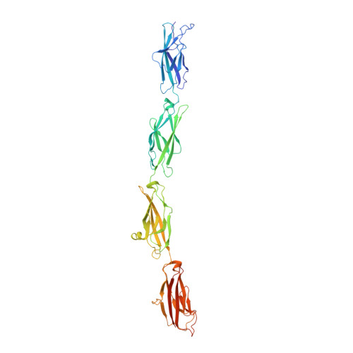
5V5X - PubMed Abstract:
Clustered protocadherins (Pcdhs) mediate numerous neural patterning functions, including neuronal self-recognition and non-self-discrimination to direct self-avoidance among vertebrate neurons. Individual neurons stochastically express a subset of Pcdh isoforms, which assemble to form a stochastic repertoire of cis -dimers. We describe the structure of a PcdhγB7 cis -homodimer, which includes the membrane-proximal extracellular cadherin domains EC5 and EC6. The structure is asymmetric with one molecule contributing interface surface from both EC5 and EC6, and the other only from EC6. Structural and sequence analyses suggest that all Pcdh isoforms will dimerize through this interface. Site-directed mutants at this interface interfere with both Pcdh cis -dimerization and cell surface transport. The structure explains the known restrictions of cis -interactions of some Pcdh isoforms, including α-Pcdhs, which cannot form homodimers. The asymmetry of the interface approximately doubles the size of the recognition repertoire, and restrictions on cis -interactions among Pcdh isoforms define the limits of the Pcdh recognition unit repertoire.
- Department of Biochemistry and Molecular Biophysics, Columbia University, New York, NY 10032.
Organizational Affiliation: