5V33

R. sphaeroides photosythetic reaction center mutant - Residue L223, Ser to Trp - Room Temperature Structure Solved on X-ray Transparent Microfluidic Chip


Experimental Data Snapshot

  • Method: X-RAY DIFFRACTION
  • Resolution: 3.49 Å
  • R-Value Free: 0.262 
  • R-Value Work: 0.221 
  • R-Value Observed: 0.225 

wwPDB Validation   3D Report Full Report


Ligand Structure Quality Assessment 


This is version 1.4 of the entry. See complete history


Literature

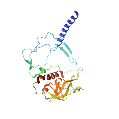
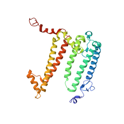
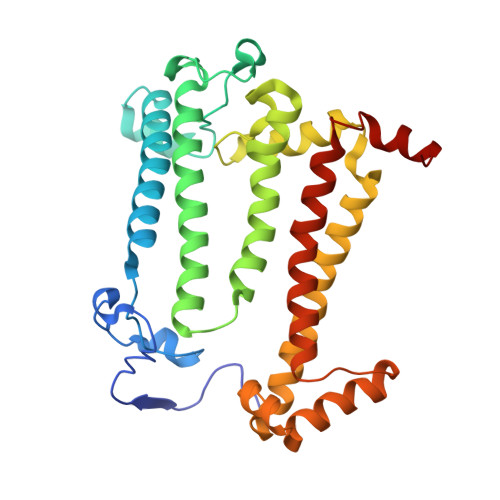
X-ray transparent microfluidic chips for high-throughput screening and optimization of in meso membrane protein crystallization.

Schieferstein, J.M.Pawate, A.S.Sun, C.Wan, F.Sheraden, P.N.Broecker, J.Ernst, O.P.Gennis, R.B.Kenis, P.J.A.

(2017) Biomicrofluidics 11: 024118-024118

  • DOI: https://doi.org/10.1063/1.4981818
  • Primary Citation of Related Structures:  
    5V33

  • PubMed Abstract: 

    Elucidating and clarifying the function of membrane proteins ultimately requires atomic resolution structures as determined most commonly by X-ray crystallography. Many high impact membrane protein structures have resulted from advanced techniques such as in meso crystallization that present technical difficulties for the set-up and scale-out of high-throughput crystallization experiments. In prior work, we designed a novel, low-throughput X-ray transparent microfluidic device that automated the mixing of protein and lipid by diffusion for in meso crystallization trials. Here, we report X-ray transparent microfluidic devices for high-throughput crystallization screening and optimization that overcome the limitations of scale and demonstrate their application to the crystallization of several membrane proteins. Two complementary chips are presented: (1) a high-throughput screening chip to test 192 crystallization conditions in parallel using as little as 8 nl of membrane protein per well and (2) a crystallization optimization chip to rapidly optimize preliminary crystallization hits through fine-gradient re-screening. We screened three membrane proteins for new in meso crystallization conditions, identifying several preliminary hits that we tested for X-ray diffraction quality. Further, we identified and optimized the crystallization condition for a photosynthetic reaction center mutant and solved its structure to a resolution of 3.5 Å.


  • Organizational Affiliation

    Department of Chemical and Biomolecular Engineering, University of Illinois at Urbana-Champaign, Urbana, Illinois 61801, USA.


Macromolecules
Find similar proteins by:  (by identity cutoff)  |  3D Structure
Entity ID: 1
MoleculeChains Sequence LengthOrganismDetailsImage
Reaction center protein H chainA [auth H]240Cereibacter sphaeroidesMutation(s): 0 
Gene Names: puhA
Membrane Entity: Yes 
UniProt
Find proteins for P0C0Y7 (Cereibacter sphaeroides)
Explore P0C0Y7 
Go to UniProtKB:  P0C0Y7
Entity Groups  
Sequence Clusters30% Identity50% Identity70% Identity90% Identity95% Identity100% Identity
UniProt GroupP0C0Y7
Sequence Annotations
Expand
  • Reference Sequence
Find similar proteins by:  (by identity cutoff)  |  3D Structure
Entity ID: 2
MoleculeChains Sequence LengthOrganismDetailsImage
Reaction center protein L chainB [auth L]281Cereibacter sphaeroidesMutation(s): 1 
Gene Names: pufL
Membrane Entity: Yes 
UniProt
Find proteins for P0C0Y8 (Cereibacter sphaeroides)
Explore P0C0Y8 
Go to UniProtKB:  P0C0Y8
Entity Groups  
Sequence Clusters30% Identity50% Identity70% Identity90% Identity95% Identity100% Identity
UniProt GroupP0C0Y8
Sequence Annotations
Expand
  • Reference Sequence
Find similar proteins by:  (by identity cutoff)  |  3D Structure
Entity ID: 3
MoleculeChains Sequence LengthOrganismDetailsImage
Reaction center protein M chainC [auth M]302Cereibacter sphaeroidesMutation(s): 0 
Gene Names: pufM
Membrane Entity: Yes 
UniProt
Find proteins for P0C0Y9 (Cereibacter sphaeroides)
Explore P0C0Y9 
Go to UniProtKB:  P0C0Y9
Entity Groups  
Sequence Clusters30% Identity50% Identity70% Identity90% Identity95% Identity100% Identity
UniProt GroupP0C0Y9
Sequence Annotations
Expand
  • Reference Sequence
Small Molecules
Ligands 4 Unique
IDChains Name / Formula / InChI Key2D Diagram3D Interactions
BCL
Query on BCL

Download Ideal Coordinates CCD File 
E [auth L],
F [auth L],
G [auth L],
J [auth M]
BACTERIOCHLOROPHYLL A
C55 H74 Mg N4 O6
DSJXIQQMORJERS-AGGZHOMASA-M
BPH
Query on BPH

Download Ideal Coordinates CCD File 
D [auth L],
K [auth M]
BACTERIOPHEOPHYTIN A
C55 H76 N4 O6
KWOZSBGNAHVCKG-SZQBJALDSA-N
U10
Query on U10

Download Ideal Coordinates CCD File 
I [auth M]UBIQUINONE-10
C59 H90 O4
ACTIUHUUMQJHFO-UPTCCGCDSA-N
FE
Query on FE

Download Ideal Coordinates CCD File 
H [auth L]FE (III) ION
Fe
VTLYFUHAOXGGBS-UHFFFAOYSA-N
Experimental Data & Validation

Experimental Data

  • Method: X-RAY DIFFRACTION
  • Resolution: 3.49 Å
  • R-Value Free: 0.262 
  • R-Value Work: 0.221 
  • R-Value Observed: 0.225 
  • Space Group: P 42 21 2
Unit Cell:
Length ( Å )Angle ( ˚ )
a = 102.332α = 90
b = 102.332β = 90
c = 240.333γ = 90
Software Package:
Software NamePurpose
PHENIXrefinement
HKL-2000data collection
HKL-2000data scaling
PDB_EXTRACTdata extraction
HKL-2000data reduction
PHASERphasing
DENZOdata reduction
SCALEPACKdata scaling

Structure Validation

View Full Validation Report


Ligand Structure Quality Assessment 


Entry History & Funding Information

Deposition Data


Funding OrganizationLocationGrant Number
National Institutes of Health/National Institute of General Medical Sciences (NIH/NIGMS)United StatesR01 GM086727

Revision History  (Full details and data files)

  • Version 1.0: 2017-04-12
    Type: Initial release
  • Version 1.1: 2017-05-17
    Changes: Database references
  • Version 1.2: 2017-09-13
    Changes: Author supporting evidence, Database references, Refinement description
  • Version 1.3: 2020-01-01
    Changes: Author supporting evidence
  • Version 1.4: 2023-10-04
    Changes: Data collection, Database references, Derived calculations, Refinement description