


1.5 A Crystal structure of PYR1 bound to Pyrabactin
Peterson, F.C., Cutler, S.R.To be published.
Experimental Data Snapshot
Starting Model: experimental
View more details
Entity ID: 1 | |||||
|---|---|---|---|---|---|
| Molecule | Chains | Sequence Length | Organism | Details | Image |
| Abscisic acid receptor PYR1 | 181 | Arabidopsis thaliana | Mutation(s): 0 Gene Names: PYR1, ABIP6, RCAR11, At4g17870, T6K21.50 | 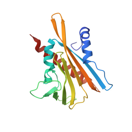 | |
UniProt | |||||
Entity Groups | |||||
| Sequence Clusters | 30% Identity50% Identity70% Identity90% Identity95% Identity100% Identity | ||||
| UniProt Group | O49686 | ||||
Sequence AnnotationsExpand | |||||
Reference Sequence | |||||
| Ligands 2 Unique | |||||
|---|---|---|---|---|---|
| ID | Chains | Name / Formula / InChI Key | 2D Diagram | 3D Interactions | |
| PYV Download:Ideal Coordinates CCD File | B [auth A] | 4-bromo-N-(pyridin-2-ylmethyl)naphthalene-1-sulfonamide C16 H13 Br N2 O2 S GJSDYQXOSHKOGX-UHFFFAOYSA-N |  | ||
| GOL Download:Ideal Coordinates CCD File | C [auth A], D [auth A], E [auth A], F [auth A], G [auth A] | GLYCEROL C3 H8 O3 PEDCQBHIVMGVHV-UHFFFAOYSA-N |  | ||
| Length ( Å ) | Angle ( ˚ ) |
|---|---|
| a = 98.427 | α = 90 |
| b = 98.427 | β = 90 |
| c = 71.467 | γ = 120 |
| Software Name | Purpose |
|---|---|
| PHENIX | refinement |
| HKL-2000 | data reduction |
| HKL-2000 | data scaling |
| PHASER | phasing |
| Funding Organization | Location | Grant Number |
|---|---|---|
| National Science Foundation (NSF, United States) | United States | 1258175 |


