Role of the Mobile Active Site Flap in IMP Dehydrogenase Inhibitor Binding.
Wang, X., Rosenberg, M.M., Kim, Y., Maltseva, N., Cuny, G.D., Joachimiak, A., Kuzmic, P., Hedstrom, L.(2025) ACS Infect Dis 11: 442-452
- PubMed: 39879323 Search on PubMedSearch on PubMed Central
- DOI: https://doi.org/10.1021/acsinfecdis.4c00636
- Primary Citation Related Structures:
5UQF, 5UQG, 5UQH, 5URQ, 5UWX, 5UXE, 5UZC, 5UZE, 5UZS, 5VSV - PubMed Abstract:
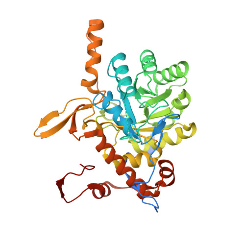
Inosine 5'-monophosphate dehydrogenase (IMPDH) is a promising antibiotic target. This enzyme catalyzes the NAD-dependent oxidation of inosine 5'-monophosphate (IMP) to xanthosine 5'-monophosphate (XMP), which is the rate-limiting step in guanine nucleotide biosynthesis. Bacterial IMPDH-specific inhibitors have been developed that bind to the NAD + site. These inhibitors display varied affinities to different bacterial IMPDHs that are not easily rationalized by X-ray crystal structures of enzyme-inhibitor complexes. Inspection of X-ray crystal structures of 25 enzyme-inhibitor complexes, including 10 newly described, suggested that a mobile active site flap may be a structural determinant of inhibitor potency. Saturation transfer difference NMR experiments also suggested that the flap may contact the inhibitors to varying extents in different IMPDHs. Flap residue Leu413 contacted some inhibitors but was not structured in the crystal structures of other inhibitor complexes. The substitution of Leu413 with Phe or Ala in Bacillus anthracis IMPDH had inhibitor-selective effects, suggesting residue 413 could be a structural determinant of affinity. Curiously, the Ala substitution increased the potency of most inhibitors, even those that contacted Leu413 in the crystal structures. Presteady-state and steady-state kinetics experiments showed that the Leu413Ala substitution had comparable effects on inhibitor binding to the noncovalent E·IMP complex and the covalent intermediate E-XMP*, suggesting that the flap had similar interactions in both complexes. These results demonstrate that contacts do not necessarily indicate favorable interactions, and poorly structured mobile regions should not be discounted when assessing binding determinants.
- Department of Chemistry, Brandeis University, Waltham, Massachusetts 02454, United States.
Organizational Affiliation: