SAR and characterization of non-substrate isoindoline urea inhibitors of nicotinamide phosphoribosyltransferase (NAMPT).
Curtin, M.L., Heyman, H.R., Clark, R.F., Sorensen, B.K., Doherty, G.A., Hansen, T.M., Frey, R.R., Sarris, K.A., Aguirre, A.L., Shrestha, A., Tu, N., Woller, K., Pliushchev, M.A., Sweis, R.F., Cheng, M., Wilsbacher, J.L., Kovar, P.J., Guo, J., Cheng, D., Longenecker, K.L., Raich, D., Korepanova, A.V., Soni, N.B., Algire, M.A., Richardson, P.L., Marin, V.L., Badagnani, I., Vasudevan, A., Buchanan, F.G., Maag, D., Chiang, G.G., Tse, C., Michaelides, M.R.(2017) Bioorg Med Chem Lett 27: 3317-3325
- PubMed: 28610984 Search on PubMed
- DOI: https://doi.org/10.1016/j.bmcl.2017.06.018
- Primary Citation Related Structures:
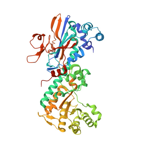
5UPE, 5UPF - PubMed Abstract:
Herein we disclose SAR studies that led to a series of isoindoline ureas which we recently reported were first-in-class, non-substrate nicotinamide phosphoribosyltransferase (NAMPT) inhibitors. Modification of the isoindoline and/or the terminal functionality of screening hit 5 provided inhibitors such as 52 and 58 with nanomolar antiproliferative activity and preclinical pharmacokinetics properties which enabled potent antitumor activity when dosed orally in mouse xenograft models. X-ray crystal structures of two inhibitors bound in the NAMPT active-site are discussed.
- AbbVie Inc, 1 North Waukegan Rd., North Chicago, IL 60064, United States. Electronic address: mike.curtin@abbvie.com.
Organizational Affiliation: